7LKY

Crystal Structure of PHF1 Tudor domain in complex with a peptidomimetic ligand UNC6641


Experimental Data Snapshot

  • Method: X-RAY DIFFRACTION
  • Resolution: 1.85 Å
  • R-Value Free: 0.281 
  • R-Value Work: 0.233 
  • R-Value Observed: 0.237 

wwPDB Validation   3D Report Full Report


This is version 1.1 of the entry. See complete history


Literature

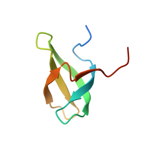
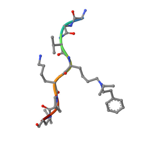
Discovery of an H3K36me3-Derived Peptidomimetic Ligand with Enhanced Affinity for Plant Homeodomain Finger Protein 1 (PHF1).

Engelberg, I.A.Liu, J.Norris-Drouin, J.L.Cholensky, S.H.Ottavi, S.A.Frye, S.V.Kutateladze, T.G.James, L.I.

(2021) J Med Chem 64: 8510-8522

  • DOI: https://doi.org/10.1021/acs.jmedchem.1c00430
  • Primary Citation of Related Structures:  
    7LKY

  • PubMed Abstract: 

    Plant homeodomain finger protein 1 (PHF1) is an accessory component of the gene silencing complex polycomb repressive complex 2 and recognizes the active chromatin mark, trimethylated lysine 36 of histone H3 (H3K36me3). In addition to its role in transcriptional regulation, PHF1 has been implicated as a driver of endometrial stromal sarcoma and fibromyxoid tumors. We report the discovery and characterization of UNC6641, a peptidomimetic antagonist of the PHF1 Tudor domain which was optimized through in silico modeling and incorporation of non-natural amino acids. UNC6641 binds the PHF1 Tudor domain with a K d value of 0.96 ± 0.03 μM while also binding the related protein PHF19 with similar potency. A crystal structure of PHF1 in complex with UNC6641, along with NMR and site-directed mutagenesis data, provided insight into the binding mechanism and requirements for binding. Additionally, UNC6641 enabled the development of a high-throughput assay to identify small molecule binders of PHF1.


  • Organizational Affiliation

    Center for Integrative Chemical Biology and Drug Discovery, Division of Chemical Biology and Medicinal Chemistry, UNC Eshelman School of Pharmacy, University of North Carolina at Chapel Hill, Chapel Hill, North Carolina 27599, United States.


Macromolecules
Find similar proteins by:  (by identity cutoff)  |  3D Structure
Entity ID: 1
MoleculeChains Sequence LengthOrganismDetailsImage
Isoform 1 of PHD finger protein 1
A, B, C, D, E
A, B, C, D, E, F, G, H
64Homo sapiensMutation(s): 0 
Gene Names: PHF1PCL1
UniProt & NIH Common Fund Data Resources
Find proteins for O43189 (Homo sapiens)
Explore O43189 
Go to UniProtKB:  O43189
PHAROS:  O43189
GTEx:  ENSG00000112511 
Entity Groups  
Sequence Clusters30% Identity50% Identity70% Identity90% Identity95% Identity100% Identity
UniProt GroupO43189
Sequence Annotations
Expand
  • Reference Sequence

Find similar proteins by:  Sequence   |   3D Structure  

Entity ID: 2
MoleculeChains Sequence LengthOrganismDetailsImage
Peptidomimetic inhibitor UNC66418synthetic constructMutation(s): 0 
Sequence Annotations
Expand
  • Reference Sequence
Small Molecules
Modified Residues  1 Unique
IDChains TypeFormula2D DiagramParent
Y57
Query on Y57
I [auth J]
J [auth I]
K
L
M
I [auth J],
J [auth I],
K,
L,
M,
N,
O,
P
L-PEPTIDE LINKINGC17 H28 N2 O2LYS
Experimental Data & Validation

Experimental Data

  • Method: X-RAY DIFFRACTION
  • Resolution: 1.85 Å
  • R-Value Free: 0.281 
  • R-Value Work: 0.233 
  • R-Value Observed: 0.237 
  • Space Group: P 1
Unit Cell:
Length ( Å )Angle ( ˚ )
a = 43.394α = 90.02
b = 45.345β = 95.19
c = 64.442γ = 90.07
Software Package:
Software NamePurpose
XSCALEdata scaling
PHENIXrefinement
PDB_EXTRACTdata extraction
autoXDSdata reduction
PHASERphasing

Structure Validation

View Full Validation Report


Entry History 

Deposition Data

Revision History  (Full details and data files)

  • Version 1.0: 2021-12-15
    Type: Initial release
  • Version 1.1: 2023-10-18
    Changes: Data collection, Refinement description