7LJM

Structure of the Salmonella enterica CD-NTase CdnD in complex with GTP


Experimental Data Snapshot

  • Method: X-RAY DIFFRACTION
  • Resolution: 2.60 Å
  • R-Value Free: 0.265 
  • R-Value Work: 0.229 
  • R-Value Observed: 0.232 

wwPDB Validation   3D Report Full Report


Ligand Structure Quality Assessment 


This is version 1.3 of the entry. See complete history


Literature

Molecular basis of CD-NTase nucleotide selection in CBASS anti-phage defense.

Govande, A.A.Duncan-Lowey, B.Eaglesham, J.B.Whiteley, A.T.Kranzusch, P.J.

(2021) Cell Rep 35: 109206-109206

  • DOI: https://doi.org/10.1016/j.celrep.2021.109206
  • Primary Citation of Related Structures:  
    7LJL, 7LJM, 7LJN, 7LJO

  • PubMed Abstract: 

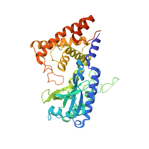
    cGAS/DncV-like nucleotidyltransferase (CD-NTase) enzymes are signaling proteins that initiate antiviral immunity in animal cells and cyclic-oligonucleotide-based anti-phage signaling system (CBASS) phage defense in bacteria. Upon phage recognition, bacterial CD-NTases catalyze synthesis of cyclic-oligonucleotide signals, which activate downstream effectors and execute cell death. How CD-NTases control nucleotide selection to specifically induce defense remains poorly defined. Here, we combine structural and nucleotide-analog interference-mapping approaches to identify molecular rules controlling CD-NTase specificity. Structures of the cyclic trinucleotide synthase Enterobacter cloacae CdnD reveal coordinating nucleotide interactions and a possible role for inverted nucleobase positioning during product synthesis. We demonstrate that correct nucleotide selection in the CD-NTase donor pocket results in the formation of a thermostable-protein-nucleotide complex, and we extend our analysis to establish specific patterns governing selectivity for each of the major bacterial CD-NTase clades A-H. Our results explain CD-NTase specificity and enable predictions of nucleotide second-messenger signals within diverse antiviral systems.


  • Organizational Affiliation

    Department of Microbiology, Harvard Medical School, Boston, MA 02115, USA; Department of Cancer Immunology and Virology, Dana-Farber Cancer Institute, Boston, MA 02115, USA.


Macromolecules
Find similar proteins by:  (by identity cutoff)  |  3D Structure
Entity ID: 1
MoleculeChains Sequence LengthOrganismDetailsImage
CD-NTase
A, B
382Salmonella entericaMutation(s): 0 
Gene Names: GNB57_001779
UniProt
Find proteins for A0A728KSL7 (Salmonella paratyphi B)
Explore A0A728KSL7 
Go to UniProtKB:  A0A728KSL7
Entity Groups  
Sequence Clusters30% Identity50% Identity70% Identity90% Identity95% Identity100% Identity
UniProt GroupA0A728KSL7
Sequence Annotations
Expand
  • Reference Sequence
Small Molecules
Ligands 2 Unique
IDChains Name / Formula / InChI Key2D Diagram3D Interactions
GTP (Subject of Investigation/LOI)
Query on GTP

Download Ideal Coordinates CCD File 
F [auth A],
G [auth A],
K [auth B],
L [auth B]
GUANOSINE-5'-TRIPHOSPHATE
C10 H16 N5 O14 P3
XKMLYUALXHKNFT-UUOKFMHZSA-N
MG
Query on MG

Download Ideal Coordinates CCD File 
C [auth A]
D [auth A]
E [auth A]
H [auth B]
I [auth B]
C [auth A],
D [auth A],
E [auth A],
H [auth B],
I [auth B],
J [auth B]
MAGNESIUM ION
Mg
JLVVSXFLKOJNIY-UHFFFAOYSA-N
Experimental Data & Validation

Experimental Data

  • Method: X-RAY DIFFRACTION
  • Resolution: 2.60 Å
  • R-Value Free: 0.265 
  • R-Value Work: 0.229 
  • R-Value Observed: 0.232 
  • Space Group: P 1 21 1
Unit Cell:
Length ( Å )Angle ( ˚ )
a = 74.473α = 90
b = 69.781β = 110.351
c = 88.697γ = 90
Software Package:
Software NamePurpose
PHENIXrefinement
XDSdata reduction
Aimlessdata scaling
AutoSolphasing

Structure Validation

View Full Validation Report


Ligand Structure Quality Assessment 


Entry History 

Deposition Data

Revision History  (Full details and data files)

  • Version 1.0: 2021-06-02
    Type: Initial release
  • Version 1.1: 2021-07-14
    Changes: Database references
  • Version 1.2: 2024-03-06
    Changes: Data collection, Database references
  • Version 1.3: 2024-04-03
    Changes: Refinement description