


Crystal Structure Analysis of human TEAD1
Seo, H.-S., Dhe-Paganon, S.To be published.
Experimental Data Snapshot
Starting Model: experimental
View more details
Entity ID: 1 | |||||
|---|---|---|---|---|---|
| Molecule | Chains | Sequence Length | Organism | Details | Image |
| Transcriptional enhancer factor TEF-1 | 220 | Homo sapiens | Mutation(s): 0 Gene Names: TEAD1 | 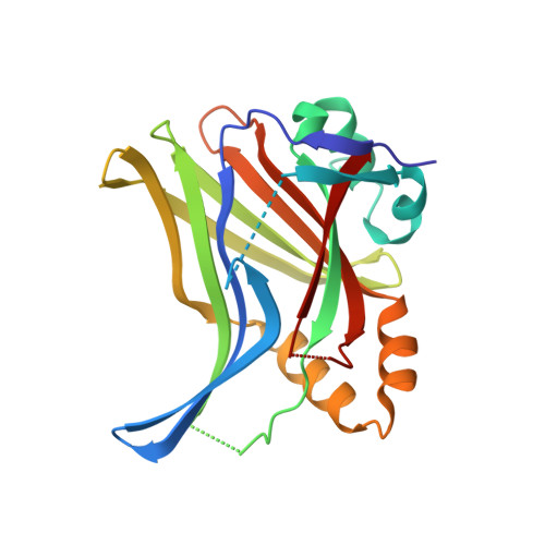 | |
UniProt & NIH Common Fund Data Resources | |||||
GTEx: ENSG00000187079 | |||||
Entity Groups | |||||
| Sequence Clusters | 30% Identity50% Identity70% Identity90% Identity95% Identity100% Identity | ||||
| UniProt Group | E9PKB7 | ||||
Sequence AnnotationsExpand | |||||
Reference Sequence | |||||
| Ligands 2 Unique | |||||
|---|---|---|---|---|---|
| ID | Chains | Name / Formula / InChI Key | 2D Diagram | 3D Interactions | |
| Y2D (Subject of Investigation/LOI) Download:Ideal Coordinates CCD File | C [auth A], E [auth B] | 1-[(3R,4R)-3-[4-(pyridin-3-yl)-1H-1,2,3-triazol-1-yl]-4-{[4-(trifluoromethyl)phenyl]methoxy}pyrrolidin-1-yl]prop-2-en-1-one C22 H20 F3 N5 O2 AZGCPBIUNCLWPF-WOJBJXKFSA-N |  | ||
| SO4 Download:Ideal Coordinates CCD File | D [auth A], F [auth B], G [auth B] | SULFATE ION O4 S QAOWNCQODCNURD-UHFFFAOYSA-L |  | ||
| Length ( Å ) | Angle ( ˚ ) |
|---|---|
| a = 36.56 | α = 90 |
| b = 89.32 | β = 90 |
| c = 135.02 | γ = 90 |
| Software Name | Purpose |
|---|---|
| PHENIX | refinement |
| xia2 | data scaling |
| PHASER | phasing |
| PDB_EXTRACT | data extraction |
| xia2 | data reduction |
| Funding Organization | Location | Grant Number |
|---|---|---|
| National Institutes of Health/National Cancer Institute (NIH/NCI) | United States | -- |


