7LHW

Structure of the LRRK2 monomer


Experimental Data Snapshot

  • Method: ELECTRON MICROSCOPY
  • Resolution: 3.70 Å
  • Aggregation State: PARTICLE 
  • Reconstruction Method: SINGLE PARTICLE 

wwPDB Validation   3D Report Full Report


This is version 1.2 of the entry. See complete history


Literature

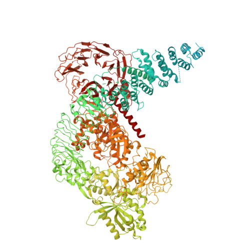
Structural analysis of the full-length human LRRK2.

Myasnikov, A.Zhu, H.Hixson, P.Xie, B.Yu, K.Pitre, A.Peng, J.Sun, J.

(2021) Cell 184: 3519

  • DOI: https://doi.org/10.1016/j.cell.2021.05.004
  • Primary Citation of Related Structures:  
    7LHT, 7LHW, 7LI3, 7LI4

  • PubMed Abstract: 

    Mutations in leucine-rich repeat kinase 2 (LRRK2) are commonly implicated in the pathogenesis of both familial and sporadic Parkinson's disease (PD). LRRK2 regulates critical cellular processes at membranous organelles and forms microtubule-based pathogenic filaments, yet the molecular basis underlying these biological roles of LRRK2 remains largely enigmatic. Here, we determined high-resolution structures of full-length human LRRK2, revealing its architecture and key interdomain scaffolding elements for rationalizing disease-causing mutations. The kinase domain of LRRK2 is captured in an inactive state, a conformation also adopted by the most common PD-associated mutation, LRRK2 G2019S . This conformation serves as a framework for structure-guided design of conformational specific inhibitors. We further determined the structure of COR-mediated LRRK2 dimers and found that single-point mutations at the dimer interface abolished pathogenic filamentation in cells. Overall, our study provides mechanistic insights into physiological and pathological roles of LRRK2 and establishes a structural template for future therapeutic intervention in PD.


  • Organizational Affiliation

    Department of Structural Biology, St. Jude Children's Research Hospital, Memphis, TN 38105, USA; Cryo-EM and Tomography Center, St. Jude Children's Research Hospital, Memphis, TN 38105, USA.


Macromolecules
Find similar proteins by:  (by identity cutoff)  |  3D Structure
Entity ID: 1
MoleculeChains Sequence LengthOrganismDetailsImage
Leucine-rich repeat serine/threonine-protein kinase 22,527Homo sapiensMutation(s): 3 
Gene Names: LRRK2PARK8
EC: 2.7.11.1 (PDB Primary Data), 3.6.5 (PDB Primary Data)
UniProt & NIH Common Fund Data Resources
Find proteins for Q5S007 (Homo sapiens)
Explore Q5S007 
Go to UniProtKB:  Q5S007
PHAROS:  Q5S007
GTEx:  ENSG00000188906 
Entity Groups  
Sequence Clusters30% Identity50% Identity70% Identity90% Identity95% Identity100% Identity
UniProt GroupQ5S007
Sequence Annotations
Expand
  • Reference Sequence
Small Molecules
Experimental Data & Validation

Experimental Data

  • Method: ELECTRON MICROSCOPY
  • Resolution: 3.70 Å
  • Aggregation State: PARTICLE 
  • Reconstruction Method: SINGLE PARTICLE 
EM Software:
TaskSoftware PackageVersion
RECONSTRUCTIONcryoSPARC3.01

Structure Validation

View Full Validation Report


Entry History & Funding Information

Deposition Data


Funding OrganizationLocationGrant Number
National Institutes of Health/National Institute on Alcohol Abuse and Alcoholism (NIH/NIAAA)United StatesHL143037(JS)
National Institutes of Health/National Institute on Alcohol Abuse and Alcoholism (NIH/NIAAA)United StatesAG053987 (JP)

Revision History  (Full details and data files)

  • Version 1.0: 2021-06-16
    Type: Initial release
  • Version 1.1: 2021-07-14
    Changes: Database references
  • Version 1.2: 2024-03-06
    Changes: Data collection, Database references