7LHU

Crystal structure of adenosine-5'-phosphosulfate reductase from Mycobacterium tuberculosis in a complex with product AMP


Experimental Data Snapshot

  • Method: X-RAY DIFFRACTION
  • Resolution: 3.09 Å
  • R-Value Free: 0.262 
  • R-Value Work: 0.230 
  • R-Value Observed: 0.231 

wwPDB Validation   3D Report Full Report


Ligand Structure Quality Assessment 


This is version 1.1 of the entry. See complete history


Literature

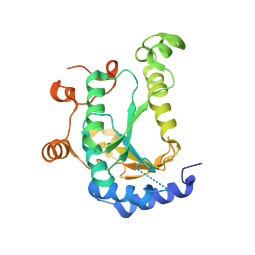
Crystal Structure of the [4Fe-4S] Cluster-Containing Adenosine-5'-phosphosulfate Reductase from Mycobacterium tuberculosis .

Feliciano, P.R.Carroll, K.S.Drennan, C.L.

(2021) ACS Omega 6: 13756-13765

  • DOI: https://doi.org/10.1021/acsomega.1c01043
  • Primary Citation of Related Structures:  
    7LHR, 7LHS, 7LHU

  • PubMed Abstract: 

    Tuberculosis (TB) is the deadliest infectious disease in the world. In Mycobacterium tuberculosis , the first committed step in sulfate assimilation is the reductive cleavage of adenosine-5'-phosphosulfate (APS) to form adenosine-5'-phosphate (AMP) and sulfite by the enzyme APS reductase (APSR). The vital role of APSR in the production of essential reduced-sulfur-containing metabolites and the absence of a homologue enzyme in humans makes APSR a potential target for therapeutic interventions. Here, we present the crystal structure of the [4Fe-4S] cluster-containing APSR from M. tuberculosis (MtbAPSR) and compare it to previously determined structures of sulfonucleotide reductases. We further present MtbAPSR structures with substrate APS and product AMP bound in the active site. Our structures at a 3.1 Å resolution show high structural similarity to other sulfonucleotide reductases and reveal that APS and AMP have similar binding modes. These studies provide structural data for structure-based drug design aimed to combat TB.


  • Organizational Affiliation

    Howard Hughes Medical Institute, Massachusetts Institute of Technology, Cambridge, Massachusetts 02139, United States.


Macromolecules
Find similar proteins by:  (by identity cutoff)  |  3D Structure
Entity ID: 1
MoleculeChains Sequence LengthOrganismDetailsImage
Phosphoadenosine phosphosulfate reductase
A, B
262Mycobacterium tuberculosisMutation(s): 0 
Gene Names: cysHMRA_2416
EC: 1.8.4.8
UniProt
Find proteins for P9WIK3 (Mycobacterium tuberculosis (strain ATCC 25618 / H37Rv))
Explore P9WIK3 
Go to UniProtKB:  P9WIK3
Entity Groups  
Sequence Clusters30% Identity50% Identity70% Identity90% Identity95% Identity100% Identity
UniProt GroupP9WIK3
Sequence Annotations
Expand
  • Reference Sequence
Experimental Data & Validation

Experimental Data

  • Method: X-RAY DIFFRACTION
  • Resolution: 3.09 Å
  • R-Value Free: 0.262 
  • R-Value Work: 0.230 
  • R-Value Observed: 0.231 
  • Space Group: P 43 21 2
Unit Cell:
Length ( Å )Angle ( ˚ )
a = 77.7α = 90
b = 77.7β = 90
c = 206.363γ = 90
Software Package:
Software NamePurpose
PHENIXrefinement
PDB_EXTRACTdata extraction
HKL-2000data reduction
HKL-2000data scaling
PHASERphasing

Structure Validation

View Full Validation Report


Ligand Structure Quality Assessment 


Entry History & Funding Information

Deposition Data


Funding OrganizationLocationGrant Number
National Institutes of Health/National Institute of General Medical Sciences (NIH/NIGMS)United StatesR35 GM126982

Revision History  (Full details and data files)

  • Version 1.0: 2021-07-07
    Type: Initial release
  • Version 1.1: 2023-10-18
    Changes: Data collection, Database references, Refinement description