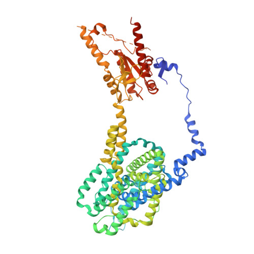
Molecular mechanism of prestin electromotive signal amplification.
Ge, J., Elferich, J., Dehghani-Ghahnaviyeh, S., Zhao, Z., Meadows, M., von Gersdorff, H., Tajkhorshid, E., Gouaux, E.(2021) Cell 184: 4669
- PubMed: 34390643 Search on PubMedSearch on PubMed Central
- DOI: https://doi.org/10.1016/j.cell.2021.07.034
- Primary Citation Related Structures:
7LGU, 7LGW, 7LH2, 7LH3 - PubMed Abstract:
Hearing involves two fundamental processes: mechano-electrical transduction and signal amplification. Despite decades of studies, the molecular bases for both remain elusive. Here, we show how prestin, the electromotive molecule of outer hair cells (OHCs) that senses both voltage and membrane tension, mediates signal amplification by coupling conformational changes to alterations in membrane surface area. Cryoelectron microscopy (cryo-EM) structures of human prestin bound with chloride or salicylate at a common "anion site" adopt contracted or expanded states, respectively. Prestin is ensconced within a perimeter of well-ordered lipids, through which it induces dramatic deformation in the membrane and couples protein conformational changes to the bulk membrane. Together with computational studies, we illustrate how the anion site is allosterically coupled to changes in the transmembrane domain cross-sectional area and the surrounding membrane. These studies provide insight into OHC electromotility by providing a structure-based mechanism of the membrane motor prestin.
- Vollum Institute, Oregon Health & Science University, 3181 SW Sam Jackson Park Road, Portland, OR 97239, USA.
Organizational Affiliation: