7LDG | pdb_00007ldg

Crystal structure of the MEILB2-BRCA2 complex

  • Classification: RECOMBINATION
  • Organism(s): Homo sapiens
  • Expression System: Escherichia coli
  • Mutation(s): No 

  • Deposited: 2021-01-13 Released: 2021-08-11 
  • Deposition Author(s): Nandakumar, J., Pendlebury, D.F.
  • Funding Organization(s): National Institutes of Health/National Institute of General Medical Sciences (NIH/NIGMS), National Institutes of Health/National Institute on Aging (NIH/NIA), American Cancer Society

Experimental Data Snapshot

  • Method: X-RAY DIFFRACTION
  • Resolution: 2.56 Å
  • R-Value Free: 
    0.231 (Depositor), 0.242 (DCC) 
  • R-Value Work: 
    0.211 (Depositor), 0.219 (DCC) 

wwPDB Validation 3D Report Full Report

Validation slider image for 7LDG

This is version 1.2 of the entry. See complete history

Literature

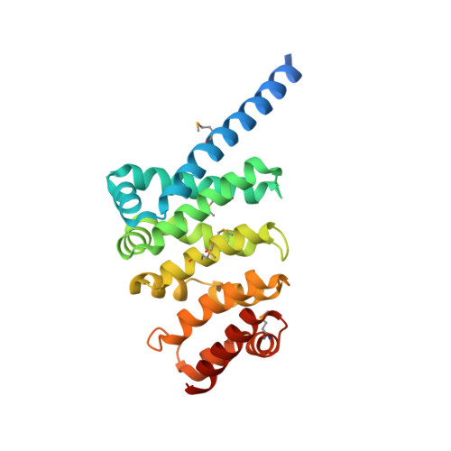
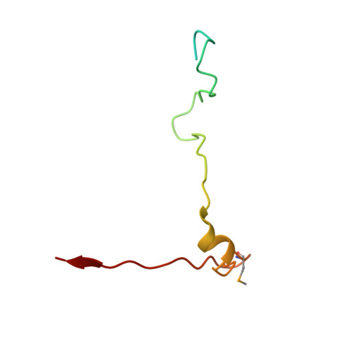
Structure of a meiosis-specific complex central to BRCA2 localization at recombination sites.

Pendlebury, D.F.Zhang, J.Agrawal, R.Shibuya, H.Nandakumar, J.

(2021) Nat Struct Mol Biol 28: 671-680

  • DOI: https://doi.org/10.1038/s41594-021-00635-0
  • Primary Citation Related Structures: 
    7LDG

  • PubMed Abstract: 

    Meiotic cells invoke breast cancer susceptibility gene 2 (BRCA2) to repair programmed double-stranded DNA breaks and accomplish homologous recombination. The meiosis-specific protein MEILB2 facilitates BRCA2 recruitment to meiotic recombination sites. Here, we combine crystallography, biochemical analysis and a mouse meiosis model to reveal a robust architecture that ensures meiotic BRCA2 recruitment. The crystal structure of the MEILB2-BRCA2 complex reveals how two MEILB2 homodimers sandwich two chains of BRCA2 to afford a 4:2 architecture. The sandwich lacks close contact between the two MEILB2 dimers or the two BRCA2 chains. Instead, the two halves of each BRCA2 chain bridge two MEILB2 subunits from different homodimers to form the MEILB2-BRCA2-MEILB2 sandwich. Several identical residues from the two MEILB2 subunits are employed to engage the BRCA2 halves, justifying their strict conservation. Mutational analysis of the interface reveals a synergistic mechanism for MEILB2-BRCA2 recruitment during meiosis. Overall, these studies demonstrate how BRCA2 efficiently localizes in the cell to facilitate meiosis.


  • Organizational Affiliation
    • Department of Molecular, Cellular, and Developmental Biology, University of Michigan, Ann Arbor, MI, USA.

Macromolecule Content 

  • Total Structure Weight: 72.09 kDa 
  • Atom Count: 3,812 
  • Modeled Residue Count: 490 
  • Deposited Residue Count: 638 
  • Unique protein chains: 2

Macromolecules

Find similar proteins by:|  3D Structure
Entity ID: 1
MoleculeChains  Sequence LengthOrganismDetailsImage
Heat shock factor 2-binding protein
A, C
253Homo sapiensMutation(s): 0 
Gene Names: HSF2BPMEILB2
UniProt & NIH Common Fund Data Resources
Find proteins for O75031 (Homo sapiens)
Explore O75031 
Go to UniProtKB:  O75031
PHAROS:  O75031
GTEx:  ENSG00000160207 
Entity Groups
Sequence Clusters30% Identity50% Identity70% Identity90% Identity95% Identity100% Identity
UniProt GroupO75031
Sequence Annotations
Expand
Reference Sequence
Find similar proteins by:|  3D Structure
Entity ID: 2
MoleculeChains  Sequence LengthOrganismDetailsImage
Breast cancer type 2 susceptibility protein
B, D
66Homo sapiensMutation(s): 0 
Gene Names: BRCA2FACDFANCD1
UniProt & NIH Common Fund Data Resources
Find proteins for P51587 (Homo sapiens)
Explore P51587 
Go to UniProtKB:  P51587
PHAROS:  P51587
GTEx:  ENSG00000139618 
Entity Groups
Sequence Clusters30% Identity50% Identity70% Identity90% Identity95% Identity100% Identity
UniProt GroupP51587
Sequence Annotations
Expand
Reference Sequence

Small Molecules

Modified Residues  1 Unique
IDChains TypeFormula2D DiagramParent
MSE
Query on MSE
A, C
L-PEPTIDE LINKINGC5 H11 N O2 SeMET

Experimental Data & Validation

Experimental Data

  • Method: X-RAY DIFFRACTION
  • Resolution: 2.56 Å
  • R-Value Free:  0.231 (Depositor), 0.242 (DCC) 
  • R-Value Work:  0.211 (Depositor), 0.219 (DCC) 
Space Group: H 3 2
Unit Cell:
Length ( Å )Angle ( ˚ )
a = 179.889α = 90
b = 179.889β = 90
c = 179.186γ = 120
Software Package:
Software NamePurpose
PHENIXrefinement
XDSdata reduction
Aimlessdata scaling
PHASERphasing

Structure Validation

View Full Validation Report


Entry History 

& Funding Information

Deposition Data


Funding OrganizationLocationGrant Number
National Institutes of Health/National Institute of General Medical Sciences (NIH/NIGMS)United StatesR01-GM120094
National Institutes of Health/National Institute on Aging (NIH/NIA)United StatesR01-AG050509
American Cancer SocietyUnited StatesRSG-17-037-01-DMC

Revision History  (Full details and data files)

  • Version 1.0: 2021-08-11
    Type: Initial release
  • Version 1.1: 2021-08-25
    Changes: Database references
  • Version 1.2: 2024-10-23
    Changes: Data collection, Structure summary