7LCL

Zoogloea ramigera biosynthetic thiolase Q183Y mutant


Experimental Data Snapshot

  • Method: X-RAY DIFFRACTION
  • Resolution: 2.29 Å
  • R-Value Free: 0.265 
  • R-Value Work: 0.229 
  • R-Value Observed: 0.231 

wwPDB Validation   3D Report Full Report


Ligand Structure Quality Assessment 


This is version 1.1 of the entry. See complete history


Literature

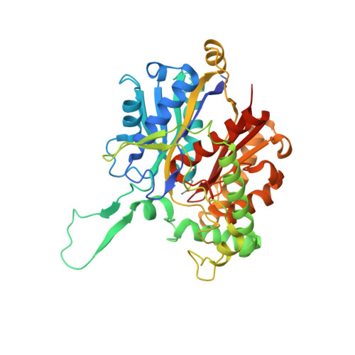
Engineering potassium activation into biosynthetic thiolase.

Marshall, A.C.Bruning, J.B.

(2021) Biochem J 478: 3047-3062

  • DOI: https://doi.org/10.1042/BCJ20210455
  • Primary Citation of Related Structures:  
    7LBZ, 7LCA, 7LCL, 7LD2, 7LDC, 7LDT, 7LDU, 7LDV, 7LDW

  • PubMed Abstract: 

    Activation of enzymes by monovalent cations (M+) is a widespread phenomenon in biology. Despite this, there are few structure-based studies describing the underlying molecular details. Thiolases are a ubiquitous and highly conserved family of enzymes containing both K+-activated and K+-independent members. Guided by structures of naturally occurring K+-activated thiolases, we have used a structure-based approach to engineer K+-activation into a K+-independent thiolase. To our knowledge, this is the first demonstration of engineering K+-activation into an enzyme, showing the malleability of proteins to accommodate M+ ions as allosteric regulators. We show that a few protein structural features encode K+-activation in this class of enzyme. Specifically, two residues near the substrate-binding site are sufficient for K+-activation: A tyrosine residue is required to complete the K+ coordination sphere, and a glutamate residue provides a compensating charge for the bound K+ ion. Further to these, a distal residue is important for positioning a K+-coordinating water molecule that forms a direct hydrogen bond to the substrate. The stability of a cation-π interaction between a positively charged residue and the substrate is determined by the conformation of the loop surrounding the substrate-binding site. Our results suggest that this cation-π interaction effectively overrides K+-activation, and is, therefore, destabilised in K+-activated thiolases. Evolutionary conservation of these amino acids provides a promising signature sequence for predicting K+-activation in thiolases. Together, our structural, biochemical and bioinformatic work provide important mechanistic insights into how enzymes can be allosterically activated by M+ ions.


  • Organizational Affiliation

    School of Molecular Sciences, The University of Western Australia, Crawley, Western Australia 6009, Australia.


Macromolecules
Find similar proteins by:  (by identity cutoff)  |  3D Structure
Entity ID: 1
MoleculeChains Sequence LengthOrganismDetailsImage
Acetyl-CoA acetyltransferase
A, B, C, D
399Zoogloea ramigeraMutation(s): 1 
Gene Names: phaAphbA
EC: 2.3.1.9
UniProt
Find proteins for P07097 (Shinella zoogloeoides)
Explore P07097 
Go to UniProtKB:  P07097
Entity Groups  
Sequence Clusters30% Identity50% Identity70% Identity90% Identity95% Identity100% Identity
UniProt GroupP07097
Sequence Annotations
Expand
  • Reference Sequence
Small Molecules
Ligands 3 Unique
IDChains Name / Formula / InChI Key2D Diagram3D Interactions
COA (Subject of Investigation/LOI)
Query on COA

Download Ideal Coordinates CCD File 
G [auth A],
N [auth B],
S [auth C],
V [auth D]
COENZYME A
C21 H36 N7 O16 P3 S
RGJOEKWQDUBAIZ-IBOSZNHHSA-N
SO4
Query on SO4

Download Ideal Coordinates CCD File 
E [auth A]
F [auth A]
H [auth A]
I [auth A]
J [auth A]
E [auth A],
F [auth A],
H [auth A],
I [auth A],
J [auth A],
L [auth B],
M [auth B],
O [auth B],
P [auth B],
Q [auth B],
T [auth C]
SULFATE ION
O4 S
QAOWNCQODCNURD-UHFFFAOYSA-L
NH4 (Subject of Investigation/LOI)
Query on NH4

Download Ideal Coordinates CCD File 
K [auth A],
R [auth B],
U [auth C],
W [auth D]
AMMONIUM ION
H4 N
QGZKDVFQNNGYKY-UHFFFAOYSA-O
Modified Residues  1 Unique
IDChains TypeFormula2D DiagramParent
SCY
Query on SCY
A, B, C, D
L-PEPTIDE LINKINGC5 H9 N O3 SCYS
Experimental Data & Validation

Experimental Data

  • Method: X-RAY DIFFRACTION
  • Resolution: 2.29 Å
  • R-Value Free: 0.265 
  • R-Value Work: 0.229 
  • R-Value Observed: 0.231 
  • Space Group: P 1 21 1
Unit Cell:
Length ( Å )Angle ( ˚ )
a = 84.175α = 90
b = 79.071β = 93.78
c = 149.635γ = 90
Software Package:
Software NamePurpose
PHENIXrefinement
XDSdata reduction
Aimlessdata scaling
PHASERphasing
PDB_EXTRACTdata extraction

Structure Validation

View Full Validation Report


Ligand Structure Quality Assessment 


Entry History 

Deposition Data

Revision History  (Full details and data files)

  • Version 1.0: 2021-09-01
    Type: Initial release
  • Version 1.1: 2023-10-18
    Changes: Data collection, Refinement description