7L6H | pdb_00007l6h

The genome-containing AAV13 capsid


Experimental Data Snapshot

  • Method: ELECTRON MICROSCOPY
  • Resolution: 3.00 Å
  • Aggregation State: PARTICLE 
  • Reconstruction Method: SINGLE PARTICLE 

wwPDB Validation 3D Report Full Report

Validation slider image for 7L6H

This is version 2.1 of the entry. See complete history

Literature

Completion of the AAV Structural Atlas: Serotype Capsid Structures Reveals Clade-Specific Features.

Mietzsch, M.Jose, A.Chipman, P.Bhattacharya, N.Daneshparvar, N.McKenna, R.Agbandje-McKenna, M.

(2021) Viruses 13

  • DOI: https://doi.org/10.3390/v13010101
  • Primary Citation Related Structures: 
    7L5Q, 7L5U, 7L6A, 7L6B, 7L6E, 7L6F, 7L6H, 7L6I

  • PubMed Abstract: 

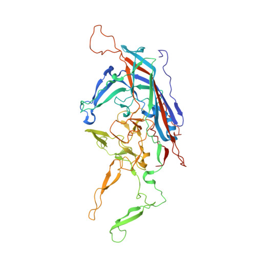
    The capsid structures of most Adeno-associated virus (AAV) serotypes, already assigned to an antigenic clade, have been previously determined. This study reports the remaining capsid structures of AAV7, AAV11, AAV12, and AAV13 determined by cryo-electron microscopy and three-dimensional image reconstruction to 2.96, 2.86, 2.54, and 2.76 Å resolution, respectively. These structures complete the structural atlas of the AAV serotype capsids. AAV7 represents the first clade D capsid structure; AAV11 and AAV12 are of a currently unassigned clade that would include AAV4; and AAV13 represents the first AAV2-AAV3 hybrid clade C capsid structure. These newly determined capsid structures all exhibit the AAV capsid features including 5-fold channels, 3-fold protrusions, 2-fold depressions, and a nucleotide binding pocket with an ordered nucleotide in genome-containing capsids. However, these structures have viral proteins that display clade-specific loop conformations. This structural characterization completes our three-dimensional library of the current AAV serotypes to provide an atlas of surface loop configurations compatible with capsid assembly and amenable for future vector engineering efforts. Derived vectors could improve gene delivery success with respect to specific tissue targeting, transduction efficiency, antigenicity or receptor retargeting.


  • Organizational Affiliation
    • Department of Biochemistry and Molecular Biology, Center for Structural Biology, McKnight Brain Institute, College of Medicine, University of Florida, Gainesville, FL 32610, USA.

Macromolecule Content 

  • Total Structure Weight: 3,533.2 kDa 
  • Atom Count: 251,400 
  • Modeled Residue Count: 31,020 
  • Deposited Residue Count: 31,020 
  • Unique protein chains: 1

Macromolecules

Find similar proteins by:|  3D Structure
Entity ID: 1
MoleculeChains  Sequence LengthOrganismDetailsImage
Capsid protein517Adeno-associated virus 13Mutation(s): 0 
UniProt
Find proteins for B5SUY7 (Adeno-associated virus 13)
Explore B5SUY7 
Go to UniProtKB:  B5SUY7
Entity Groups
Sequence Clusters30% Identity50% Identity70% Identity90% Identity95% Identity100% Identity
UniProt GroupB5SUY7
Sequence Annotations
Expand
Reference Sequence

Small Molecules

Ligands 1 Unique
IDChains Name / Formula / InChI Key2D Diagram3D Interactions
D5M

Query on D5M


Download:Ideal Coordinates CCD File
AC [auth S]
AD [auth s]
BC [auth T]
BD [auth t]
CC [auth U]
AC [auth S],
AD [auth s],
BC [auth T],
BD [auth t],
CC [auth U],
CD [auth u],
DC [auth V],
DD [auth v],
EC [auth W],
ED [auth w],
FC [auth X],
FD [auth x],
GC [auth Y],
GD [auth y],
HC [auth Z],
HD [auth z],
IB [auth A],
IC [auth a],
ID [auth 1],
JB [auth B],
JC [auth b],
JD [auth 2],
KB [auth C],
KC [auth c],
KD [auth 3],
LB [auth D],
LC [auth d],
LD [auth 4],
MB [auth E],
MC [auth e],
MD [auth 5],
NB [auth F],
NC [auth f],
ND [auth 6],
OB [auth G],
OC [auth g],
OD [auth 7],
PB [auth H],
PC [auth h],
PD [auth 8],
QB [auth I],
QC [auth i],
RB [auth J],
RC [auth j],
SB [auth K],
SC [auth k],
TB [auth L],
TC [auth l],
UB [auth M],
UC [auth m],
VB [auth N],
VC [auth n],
WB [auth O],
WC [auth o],
XB [auth P],
XC [auth p],
YB [auth Q],
YC [auth q],
ZB [auth R],
ZC [auth r]
2'-DEOXYADENOSINE-5'-MONOPHOSPHATE
C10 H14 N5 O6 P
KHWCHTKSEGGWEX-RRKCRQDMSA-N

Experimental Data & Validation

Experimental Data

  • Method: ELECTRON MICROSCOPY
  • Resolution: 3.00 Å
  • Aggregation State: PARTICLE 
  • Reconstruction Method: SINGLE PARTICLE 
EM Software:
TaskSoftware PackageVersion
MODEL REFINEMENTPHENIX
RECONSTRUCTIONcisTEM

Structure Validation

View Full Validation Report


Entry History 

& Funding Information

Deposition Data


Funding OrganizationLocationGrant Number
National Institutes of Health/National Institute of General Medical Sciences (NIH/NIGMS)United StatesGM082946

Revision History  (Full details and data files)

  • Version 1.0: 2021-02-03
    Type: Initial release
  • Version 1.1: 2024-03-06
    Changes: Data collection, Database references
  • Version 2.0: 2024-05-29
    Changes: Atomic model, Data collection, Derived calculations, Non-polymer description
  • Version 2.1: 2025-05-28
    Changes: Data collection, Structure summary