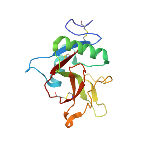
Structural analysis of carbohydrate binding by the macrophage mannose receptor CD206.
Feinberg, H., Jegouzo, S.A.F., Lasanajak, Y., Smith, D.F., Drickamer, K., Weis, W.I., Taylor, M.E.(2021) J Biological Chem 296: 100368-100368
- PubMed: 33545173 Search on PubMedSearch on PubMed Central
- DOI: https://doi.org/10.1016/j.jbc.2021.100368
- Primary Citation Related Structures:
7JUB, 7JUC, 7JUD, 7JUE, 7JUF, 7JUG, 7JUH, 7L61, 7L62, 7L63, 7L64, 7L65, 7L66, 7L67, 7L68 - PubMed Abstract:
The human mannose receptor expressed on macrophages and hepatic endothelial cells scavenges released lysosomal enzymes, glycopeptide fragments of collagen, and pathogenic microorganisms and thus reduces damage following tissue injury. The receptor binds mannose, fucose, or N-acetylglucosamine (GlcNAc) residues on these targets. C-type carbohydrate-recognition domain 4 (CRD4) of the receptor contains the site for Ca 2+ -dependent interaction with sugars. To investigate the details of CRD4 binding, glycan array screening was used to identify oligosaccharide ligands. The strongest signals were for glycans that contain either Manα1-2Man constituents or fucose in various linkages. The mechanisms of binding to monosaccharides and oligosaccharide substructures present in many of these ligands were examined in multiple crystal structures of CRD4. Binding of mannose residues to CRD4 results primarily from interaction of the equatorial 3- and 4-OH groups with a conserved principal Ca 2+ common to almost all sugar-binding C-type CRDs. In the Manα1-2Man complex, supplementary interactions with the reducing mannose residue explain the enhanced affinity for this disaccharide. Bound GlcNAc also interacts with the principal Ca 2+ through equatorial 3- and 4-OH groups, whereas fucose residues can bind in several orientations, through either the 2- and 3-OH groups or the 3- and 4-OH groups. Secondary contacts with additional sugars in fucose-containing oligosaccharides, such as the Lewis-a trisaccharide, provide enhanced affinity for these glycans. These results explain many of the biologically important interactions of the mannose receptor with both mammalian glycoproteins and microbes such as yeast and suggest additional classes of ligands that have not been previously identified.
- Departments of Structural Biology and Molecular and Cellular Physiology, Stanford University School of Medicine, Stanford, California, USA.
Organizational Affiliation: