7L0Z | pdb_00007l0z

Spinach variant bound to DFHBI-1T


Experimental Data Snapshot

  • Method: X-RAY DIFFRACTION
  • Resolution: 2.10 Å
  • R-Value Free: 
    0.232 (Depositor), 0.234 (DCC) 
  • R-Value Work: 
    0.196 (Depositor), 0.199 (DCC) 
  • R-Value Observed: 
    0.200 (Depositor) 

Starting Model: experimental
View more details

wwPDB Validation 3D Report Full Report

Validation slider image for 7L0Z

Ligand Structure Quality Assessment 


This is version 1.1 of the entry. See complete history

Literature

Fluorogenic aptamers resolve the flexibility of RNA junctions using orientation-dependent FRET.

Jeng, S.C.Y.Trachman III, R.J.Weissenboeck, F.Truong, L.Link, K.A.Jepsen, M.D.E.Knutson, J.R.Andersen, E.S.Ferre-D'Amare, A.R.Unrau, P.J.

(2021) RNA 27: 433-444

  • DOI: https://doi.org/10.1261/rna.078220.120
  • Primary Citation Related Structures: 
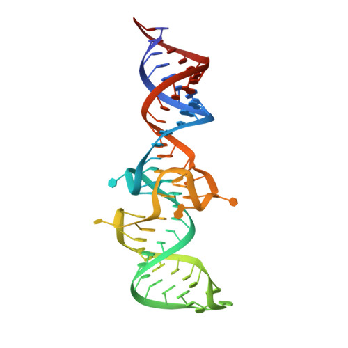
    6UP0, 7L0Z

  • PubMed Abstract: 

    To further understand the transcriptome, new tools capable of measuring folding, interactions, and localization of RNA are needed. Although Förster resonance energy transfer (FRET) is an angle- and distance-dependent phenomenon, the majority of FRET measurements have been used to report distances, by assuming rotationally averaged donor-acceptor pairs. Angle-dependent FRET measurements have proven challenging for nucleic acids due to the difficulties in incorporating fluorophores rigidly into local substructures in a biocompatible manner. Fluorescence turn-on RNA aptamers are genetically encodable tags that appear to rigidly confine their cognate fluorophores, and thus have the potential to report angular-resolved FRET. Here, we use the fluorescent aptamers Broccoli and Mango-III as donor and acceptor, respectively, to measure the angular dependence of FRET. Joining the two fluorescent aptamers by a helix of variable length allowed systematic rotation of the acceptor fluorophore relative to the donor. FRET oscillated in a sinusoidal manner as a function of helix length, consistent with simulated data generated from models of oriented fluorophores separated by an inflexible helix. Analysis of the orientation dependence of FRET allowed us to demonstrate structural rigidification of the NiCo riboswitch upon transition metal-ion binding. This application of fluorescence turn-on aptamers opens the way to improved structural interpretation of ensemble and single-molecule FRET measurements of RNA.


  • Organizational Affiliation
    • Department of Molecular Biology and Biochemistry, Simon Fraser University, Burnaby, British Columbia, Canada V5A 1S6.

Macromolecule Content 

  • Total Structure Weight: 23.64 kDa 
  • Atom Count: 1,612 
  • Modeled Residue Count: 69 
  • Deposited Residue Count: 69 
  • Unique nucleic acid chains: 1

Macromolecules

Find similar nucleic acids by:  (by identity cutoff) 
Entity ID: 1
MoleculeChains LengthOrganismImage
RNA (69-MER)A [auth G]69synthetic construct
Sequence Annotations
Expand
Reference Sequence

Small Molecules

Ligands 6 Unique
IDChains Name / Formula / InChI Key2D Diagram3D Interactions
2ZY

Query on 2ZY


Download:Ideal Coordinates CCD File
B [auth G](5Z)-5-(3,5-difluoro-4-hydroxybenzylidene)-2-methyl-3-(2,2,2-trifluoroethyl)-3,5-dihydro-4H-imidazol-4-one
C13 H9 F5 N2 O2
AWYCLBWNRONMQC-WMZJFQQLSA-N
SPM

Query on SPM


Download:Ideal Coordinates CCD File
C [auth G],
D [auth G],
E [auth G]
SPERMINE
C10 H26 N4
PFNFFQXMRSDOHW-UHFFFAOYSA-N
PO4

Query on PO4


Download:Ideal Coordinates CCD File
G,
H [auth G]
PHOSPHATE ION
O4 P
NBIIXXVUZAFLBC-UHFFFAOYSA-K
K

Query on K


Download:Ideal Coordinates CCD File
K [auth G],
L [auth G]
POTASSIUM ION
K
NPYPAHLBTDXSSS-UHFFFAOYSA-N
MG

Query on MG


Download:Ideal Coordinates CCD File
F [auth G]MAGNESIUM ION
Mg
JLVVSXFLKOJNIY-UHFFFAOYSA-N
NA

Query on NA


Download:Ideal Coordinates CCD File
I [auth G],
J [auth G]
SODIUM ION
Na
FKNQFGJONOIPTF-UHFFFAOYSA-N

Experimental Data & Validation

Experimental Data

  • Method: X-RAY DIFFRACTION
  • Resolution: 2.10 Å
  • R-Value Free:  0.232 (Depositor), 0.234 (DCC) 
  • R-Value Work:  0.196 (Depositor), 0.199 (DCC) 
  • R-Value Observed: 0.200 (Depositor) 
Space Group: C 1 2 1
Unit Cell:
Length ( Å )Angle ( ˚ )
a = 150.564α = 90
b = 48.692β = 91.364
c = 30.612γ = 90
Software Package:
Software NamePurpose
PHENIXrefinement
XDSdata reduction
XDSdata scaling
PHASERphasing

Structure Validation

View Full Validation Report


Ligand Structure Quality Assessment 


Entry History 

Deposition Data

Revision History  (Full details and data files)

  • Version 1.0: 2021-05-26
    Type: Initial release
  • Version 1.1: 2023-10-18
    Changes: Data collection, Database references, Refinement description