7KZL | pdb_00007kzl

Cyclopentane peptide nucleic acid in complex with DNA


Experimental Data Snapshot

  • Method: X-RAY DIFFRACTION
  • Resolution: 1.30 Å
  • R-Value Free: 
    0.189 (Depositor), 0.197 (DCC) 
  • R-Value Work: 
    0.172 (Depositor), 0.178 (DCC) 
  • R-Value Observed: 
    0.173 (Depositor) 

Starting Model: experimental
View more details

wwPDB Validation 3D Report Full Report

Validation slider image for 7KZL

This is version 2.2 of the entry. See complete history

Literature

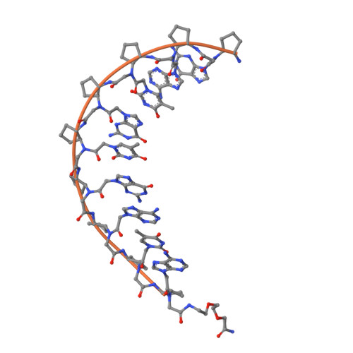
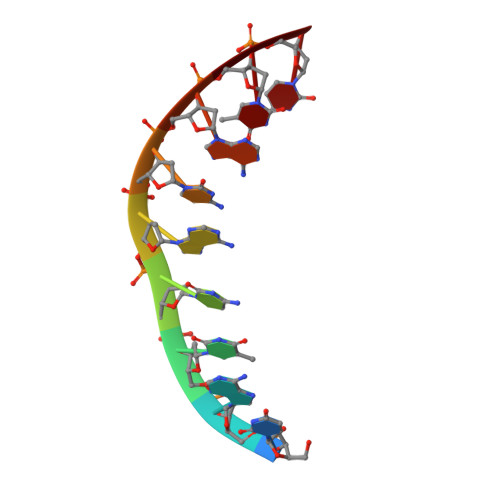
Conformational constraints of cyclopentane peptide nucleic acids facilitate tunable binding to DNA.

Zheng, H.Botos, I.Clausse, V.Nikolayevskiy, H.Rastede, E.E.Fouz, M.F.Mazur, S.J.Appella, D.H.

(2021) Nucleic Acids Res 49: 713-725

  • DOI: https://doi.org/10.1093/nar/gkaa1249
  • Primary Citation Related Structures: 
    7KZL

  • PubMed Abstract: 

    We report a series of synthetic, nucleic acid mimics with highly customizable thermodynamic binding to DNA. Incorporation of helix-promoting cyclopentanes into peptide nucleic acids (PNAs) increases the melting temperatures (Tm) of PNA+DNA duplexes by approximately +5°C per cyclopentane. Sequential addition of cyclopentanes allows the Tm of PNA + DNA duplexes to be systematically fine-tuned from +5 to +50°C compared with the unmodified PNA. Containing only nine nucleobases and an equal number of cyclopentanes, cpPNA-9 binds to complementary DNA with a Tm around 90°C. Additional experiments reveal that the cpPNA-9 sequence specifically binds to DNA duplexes containing its complementary sequence and functions as a PCR clamp. An X-ray crystal structure of the cpPNA-9-DNA duplex revealed that cyclopentanes likely induce a right-handed helix in the PNA with conformations that promote DNA binding.


  • Organizational Affiliation
    • Synthetic Bioactive Molecules Section, Laboratory of Bioorganic Chemistry (LBC), National Institute of Diabetes and Digestive and Kidney Diseases, National Institutes of Health, 8 Center Drive, Room 404, Bethesda, MD 20892, USA.

Macromolecule Content 

  • Total Structure Weight: 6.37 kDa 
  • Atom Count: 508 
  • Modeled Residue Count: 19 
  • Deposited Residue Count: 19 
  • Unique protein chains: 1
  • Unique nucleic acid chains: 1

Macromolecules


Find similar proteins by:  Sequence   |   3D Structure  
Entity ID: 2
MoleculeChains  Sequence LengthOrganismDetailsImage
XDJ-XDD-XDY-XDJ-XDY-XDJ-XDD-XDY-XDD-XDV10synthetic constructMutation(s): 0 
Find similar nucleic acids by:  Sequence
Entity ID: 1
MoleculeChains LengthOrganismImage
DNA (5'-D(*TP*AP*TP*CP*AP*CP*AP*TP*C)-3')9synthetic construct
Sequence Annotations
Expand
Reference Sequence

Small Molecules

Ligands 4 Unique
IDChains Name / Formula / InChI Key2D Diagram3D Interactions
IOD

Query on IOD


Download:Ideal Coordinates CCD File
C [auth A]IODIDE ION
I
XMBWDFGMSWQBCA-UHFFFAOYSA-M
ZN

Query on ZN


Download:Ideal Coordinates CCD File
D [auth A]
E [auth A]
J [auth B]
K [auth B]
L [auth B]
D [auth A],
E [auth A],
J [auth B],
K [auth B],
L [auth B],
M [auth B]
ZINC ION
Zn
PTFCDOFLOPIGGS-UHFFFAOYSA-N
EDO

Query on EDO


Download:Ideal Coordinates CCD File
I [auth B]1,2-ETHANEDIOL
C2 H6 O2
LYCAIKOWRPUZTN-UHFFFAOYSA-N
MOH

Query on MOH


Download:Ideal Coordinates CCD File
F [auth B],
G [auth B],
H [auth B]
METHANOL
C H4 O
OKKJLVBELUTLKV-UHFFFAOYSA-N

Experimental Data & Validation

Experimental Data

  • Method: X-RAY DIFFRACTION
  • Resolution: 1.30 Å
  • R-Value Free:  0.189 (Depositor), 0.197 (DCC) 
  • R-Value Work:  0.172 (Depositor), 0.178 (DCC) 
  • R-Value Observed: 0.173 (Depositor) 
Space Group: P 43 21 2
Unit Cell:
Length ( Å )Angle ( ˚ )
a = 54.704α = 90
b = 54.704β = 90
c = 39.633γ = 90
Software Package:
Software NamePurpose
PHENIXrefinement
HKL-2000data reduction
HKL-2000data scaling
PHENIXphasing

Structure Validation

View Full Validation Report


Entry History 

& Funding Information

Deposition Data


Funding OrganizationLocationGrant Number
National Institutes of Health/National Institute of Diabetes and Digestive and Kidney Disease (NIH/NIDDK)United StatesZIA-DK031143

Revision History  (Full details and data files)

  • Version 1.0: 2020-12-23
    Type: Initial release
  • Version 1.1: 2021-01-20
    Changes: Database references
  • Version 1.2: 2021-02-03
    Changes: Database references
  • Version 2.0: 2023-11-15
    Changes: Atomic model, Data collection, Database references, Derived calculations
  • Version 2.1: 2024-04-03
    Changes: Refinement description
  • Version 2.2: 2024-10-09
    Changes: Structure summary