7KWZ | pdb_00007kwz

TDP-43 LCD amyloid fibrils

  • Classification: PROTEIN FIBRIL
  • Organism(s): Homo sapiens
  • Expression System: Escherichia coli
  • Mutation(s): No 

  • Deposited: 2020-12-02 Released: 2021-02-24 
  • Deposition Author(s): Li, Q., Babinchak, W.M., Surewicz, W.K.
  • Funding Organization(s): National Institutes of Health/National Institute on Aging (NIH/NIA), National Institutes of Health/National Institute of General Medical Sciences (NIH/NIGMS)

Experimental Data Snapshot

  • Method: ELECTRON MICROSCOPY
  • Resolution: 3.20 Å
  • Aggregation State: FILAMENT 
  • Reconstruction Method: HELICAL 

wwPDB Validation 3D Report Full Report

Validation slider image for 7KWZ

This is version 1.2 of the entry. See complete history

Literature

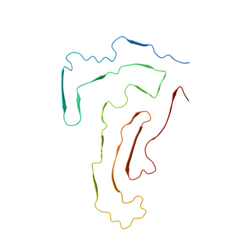
Cryo-EM structure of amyloid fibrils formed by the entire low complexity domain of TDP-43.

Li, Q.Babinchak, W.M.Surewicz, W.K.

(2021) Nat Commun 12: 1620-1620

  • DOI: https://doi.org/10.1038/s41467-021-21912-y
  • Primary Citation Related Structures: 
    7KWZ

  • PubMed Abstract: 

    Amyotrophic lateral sclerosis and several other neurodegenerative diseases are associated with brain deposits of amyloid-like aggregates formed by the C-terminal fragments of TDP-43 that contain the low complexity domain of the protein. Here, we report the cryo-EM structure of amyloid formed from the entire TDP-43 low complexity domain in vitro at pH 4. This structure reveals single protofilament fibrils containing a large (139-residue), tightly packed core. While the C-terminal part of this core region is largely planar and characterized by a small proportion of hydrophobic amino acids, the N-terminal region contains numerous hydrophobic residues and has a non-planar backbone conformation, resulting in rugged surfaces of fibril ends. The structural features found in these fibrils differ from those previously found for fibrils generated from short protein fragments. The present atomic model for TDP-43 LCD fibrils provides insight into potential structural perturbations caused by phosphorylation and disease-related mutations.


  • Organizational Affiliation
    • Department of Physiology and Biophysics, Case Western Reserve University, Cleveland, OH, USA.

Macromolecule Content 

  • Total Structure Weight: 72.85 kDa 
  • Atom Count: 4,695 
  • Modeled Residue Count: 695 
  • Deposited Residue Count: 740 
  • Unique protein chains: 1

Macromolecules

Find similar proteins by:|  3D Structure
Entity ID: 1
MoleculeChains  Sequence LengthOrganismDetailsImage
Isoform 2 of TAR DNA-binding protein 43
A, B, C, D, E
148Homo sapiensMutation(s): 0 
Gene Names: TARDBPTDP43
UniProt & NIH Common Fund Data Resources
Find proteins for Q13148 (Homo sapiens)
Explore Q13148 
Go to UniProtKB:  Q13148
PHAROS:  Q13148
GTEx:  ENSG00000120948 
Entity Groups
Sequence Clusters30% Identity50% Identity70% Identity90% Identity95% Identity100% Identity
UniProt GroupQ13148
Sequence Annotations
Expand
Reference Sequence

Experimental Data & Validation

Experimental Data

  • Method: ELECTRON MICROSCOPY
  • Resolution: 3.20 Å
  • Aggregation State: FILAMENT 
  • Reconstruction Method: HELICAL 

Structure Validation

View Full Validation Report


Entry History 

& Funding Information

Deposition Data


Funding OrganizationLocationGrant Number
National Institutes of Health/National Institute on Aging (NIH/NIA)United StatesAG061797
National Institutes of Health/National Institute of General Medical Sciences (NIH/NIGMS)United StatesGM094357

Revision History  (Full details and data files)

  • Version 1.0: 2021-02-24
    Type: Initial release
  • Version 1.1: 2021-03-24
    Changes: Database references
  • Version 1.2: 2024-03-06
    Changes: Data collection, Database references, Derived calculations