7KUQ

Crystal Structure of Danio rerio Histone Deacetylase 10 Y307F Mutant in Complex with N8-Acetylspermidine


Experimental Data Snapshot

  • Method: X-RAY DIFFRACTION
  • Resolution: 2.10 Å
  • R-Value Free: 0.247 
  • R-Value Work: 0.205 
  • R-Value Observed: 0.207 

wwPDB Validation   3D Report Full Report


Ligand Structure Quality Assessment 


This is version 1.2 of the entry. See complete history


Literature

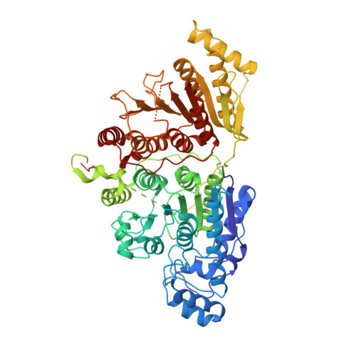
X-ray Crystallographic Snapshots of Substrate Binding in the Active Site of Histone Deacetylase 10.

Herbst-Gervasoni, C.J.Christianson, D.W.

(2021) Biochemistry 60: 303-313

  • DOI: https://doi.org/10.1021/acs.biochem.0c00936
  • Primary Citation of Related Structures:  
    7KUQ, 7KUR, 7KUS, 7KUT, 7KUV

  • PubMed Abstract: 

    Histone deacetylase 10 (HDAC10) is a zinc-dependent polyamine deacetylase enriched in the cytosol of eukaryotic cells. The active site of HDAC10 contains catalytic residues conserved in other HDAC isozymes that function as lysine deacetylases: Y307 assists the zinc ion in polarizing the substrate carbonyl for nucleophilic attack, and the H136-H137 dyad serves general base-general acid functions. As an inducer of autophagy, HDAC10 is an attractive target for the design of selective inhibitors that may be useful in cancer chemotherapy. Because detailed structural information regarding the catalytic mechanism of HDAC10 may inform new approaches to inhibitor design, we now report X-ray crystal structures of HDAC10 in which reaction intermediates with substrates N 8 -acetylspermidine and N -acetylputrescine are trapped in the active site. The Y307F substitution prevents activation of the substrate carbonyl for nucleophilic attack by the zinc-bound water molecule, thereby enabling crystallographic isolation of intact enzyme-substrate complexes. The H137A substitution removes the catalytically obligatory general acid, thereby enabling crystallographic isolation of oxyanionic tetrahedral intermediates. Finally, the acetate complex with the wild-type enzyme represents a product complex after dissociation of the polyamine coproduct. Taken together, these structures provide snapshots of the reaction coordinate of acetylpolyamine hydrolysis and are consistent with a mechanism in which tandem histidine residues H136 and H137 serve as general base and general acid catalysts, respectively. The function of the histidine dyad in the HDAC10 mechanism appears to be similar to that in HDAC6, but not HDAC8 in which both functions are served by the second histidine of the tandem pair.


  • Organizational Affiliation

    Roy and Diana Vagelos Laboratories, Department of Chemistry, University of Pennsylvania, Philadelphia, Pennsylvania 19104-6323, United States.


Macromolecules
Find similar proteins by:  (by identity cutoff)  |  3D Structure
Entity ID: 1
MoleculeChains Sequence LengthOrganismDetailsImage
Polyamine deacetylase HDAC10678Danio rerioMutation(s): 1 
Gene Names: hdac10
EC: 3.5.1.48 (PDB Primary Data), 3.5.1.62 (PDB Primary Data)
UniProt
Find proteins for F1QCV2 (Danio rerio)
Explore F1QCV2 
Go to UniProtKB:  F1QCV2
Entity Groups  
Sequence Clusters30% Identity50% Identity70% Identity90% Identity95% Identity100% Identity
UniProt GroupF1QCV2
Sequence Annotations
Expand
  • Reference Sequence
Small Molecules
Ligands 5 Unique
IDChains Name / Formula / InChI Key2D Diagram3D Interactions
Q9C (Subject of Investigation/LOI)
Query on Q9C

Download Ideal Coordinates CCD File 
B [auth A]N-{4-[(3-aminopropyl)amino]butyl}acetamide
C9 H21 N3 O
FONIWJIDLJEJTL-UHFFFAOYSA-N
PO4
Query on PO4

Download Ideal Coordinates CCD File 
F [auth A],
G [auth A],
H [auth A]
PHOSPHATE ION
O4 P
NBIIXXVUZAFLBC-UHFFFAOYSA-K
ZN
Query on ZN

Download Ideal Coordinates CCD File 
C [auth A]ZINC ION
Zn
PTFCDOFLOPIGGS-UHFFFAOYSA-N
K
Query on K

Download Ideal Coordinates CCD File 
D [auth A],
E [auth A]
POTASSIUM ION
K
NPYPAHLBTDXSSS-UHFFFAOYSA-N
NA
Query on NA

Download Ideal Coordinates CCD File 
I [auth A]SODIUM ION
Na
FKNQFGJONOIPTF-UHFFFAOYSA-N
Experimental Data & Validation

Experimental Data

  • Method: X-RAY DIFFRACTION
  • Resolution: 2.10 Å
  • R-Value Free: 0.247 
  • R-Value Work: 0.205 
  • R-Value Observed: 0.207 
  • Space Group: P 31 2 1
Unit Cell:
Length ( Å )Angle ( ˚ )
a = 80.509α = 90
b = 80.509β = 90
c = 249.38γ = 120
Software Package:
Software NamePurpose
PHENIXrefinement
iMOSFLMdata reduction
Aimlessdata scaling
PHASERphasing

Structure Validation

View Full Validation Report


Ligand Structure Quality Assessment 


Entry History & Funding Information

Deposition Data


Funding OrganizationLocationGrant Number
National Institutes of Health/National Human Genome Research Institute (NIH/NHGRI)United StatesGM49758

Revision History  (Full details and data files)

  • Version 1.0: 2021-02-03
    Type: Initial release
  • Version 1.1: 2021-02-17
    Changes: Database references
  • Version 1.2: 2023-10-18
    Changes: Data collection, Database references, Refinement description