7KQ8 | pdb_00007kq8

Structure of iron bound MEMO1


Experimental Data Snapshot

  • Method: X-RAY DIFFRACTION
  • Resolution: 2.15 Å
  • R-Value Free: 
    0.202 (Depositor), 0.203 (DCC) 
  • R-Value Work: 
    0.159 (Depositor), 0.161 (DCC) 
  • R-Value Observed: 
    0.160 (Depositor) 

Starting Model: experimental
View more details

wwPDB Validation 3D Report Full Report

Validation slider image for 7KQ8

This is version 1.2 of the entry. See complete history

Literature

MEMO1 binds iron and modulates iron homeostasis in cancer cells.

Dolgova, N.Uhlemann, E.E.Boniecki, M.T.Vizeacoumar, F.S.Ara, A.Nouri, P.Ralle, M.Tonelli, M.Abbas, S.A.Patry, J.Elhasasna, H.Freywald, A.Vizeacoumar, F.J.Dmitriev, O.Y.

(2024) Elife 13

  • DOI: https://doi.org/10.7554/eLife.86354
  • Primary Citation Related Structures: 
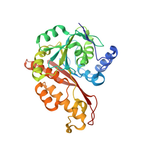
    7KQ8, 7L5C, 7M8H

  • PubMed Abstract: 

    Mediator of ERBB2-driven cell motility 1 (MEMO1) is an evolutionary conserved protein implicated in many biological processes; however, its primary molecular function remains unknown. Importantly, MEMO1 is overexpressed in many types of cancer and was shown to modulate breast cancer metastasis through altered cell motility. To better understand the function of MEMO1 in cancer cells, we analyzed genetic interactions of MEMO1 using gene essentiality data from 1028 cancer cell lines and found multiple iron-related genes exhibiting genetic relationships with MEMO1. We experimentally confirmed several interactions between MEMO1 and iron-related proteins in living cells, most notably, transferrin receptor 2 ( TFR 2), mitoferrin-2 ( SLC25A28 ), and the global iron response regulator IRP1 ( ACO1 ). These interactions indicate that cells with high-MEMO1 expression levels are hypersensitive to the disruptions in iron distribution. Our data also indicate that MEMO1 is involved in ferroptosis and is linked to iron supply to mitochondria. We have found that purified MEMO1 binds iron with high affinity under redox conditions mimicking intracellular environment and solved MEMO1 structures in complex with iron and copper. Our work reveals that the iron coordination mode in MEMO1 is very similar to that of iron-containing extradiol dioxygenases, which also display a similar structural fold. We conclude that MEMO1 is an iron-binding protein that modulates iron homeostasis in cancer cells.


  • Organizational Affiliation
    • Department of Biochemistry, Microbiology and Immunology, University of Saskatchewan, Saskatoon, Canada.

Macromolecule Content 

  • Total Structure Weight: 138.42 kDa 
  • Atom Count: 10,317 
  • Modeled Residue Count: 1,176 
  • Deposited Residue Count: 1,176 
  • Unique protein chains: 1

Macromolecules

Find similar proteins by:|  3D Structure
Entity ID: 1
MoleculeChains  Sequence LengthOrganismDetailsImage
Protein MEMO1
A, B, C, D
294Homo sapiensMutation(s): 0 
Gene Names: MEMO1C2orf4MEMONS5ATP7CGI-27
UniProt & NIH Common Fund Data Resources
Find proteins for Q9Y316 (Homo sapiens)
Explore Q9Y316 
Go to UniProtKB:  Q9Y316
PHAROS:  Q9Y316
GTEx:  ENSG00000162959 
Entity Groups
Sequence Clusters30% Identity50% Identity70% Identity90% Identity95% Identity100% Identity
UniProt GroupQ9Y316
Sequence Annotations
Expand
Reference Sequence

Small Molecules

Ligands 5 Unique
IDChains Name / Formula / InChI Key2D Diagram3D Interactions
GSH

Query on GSH


Download:Ideal Coordinates CCD File
E [auth A],
LA [auth C],
V [auth B],
WA [auth D]
Glutathione
C10 H17 N3 O6 S
RWSXRVCMGQZWBV-WDSKDSINSA-N
PEG

Query on PEG


Download:Ideal Coordinates CCD File
GA [auth B],
PA [auth C],
QA [auth C]
DI(HYDROXYETHYL)ETHER
C4 H10 O3
MTHSVFCYNBDYFN-UHFFFAOYSA-N
GOL

Query on GOL


Download:Ideal Coordinates CCD File
RA [auth C],
W [auth B],
XA [auth D]
GLYCEROL
C3 H8 O3
PEDCQBHIVMGVHV-UHFFFAOYSA-N
EDO

Query on EDO


Download:Ideal Coordinates CCD File
AA [auth B]
AB [auth D]
BA [auth B]
BB [auth D]
CA [auth B]
AA [auth B],
AB [auth D],
BA [auth B],
BB [auth D],
CA [auth B],
CB [auth D],
DA [auth B],
DB [auth D],
EA [auth B],
EB [auth D],
F [auth A],
FA [auth B],
FB [auth D],
G [auth A],
GB [auth D],
H [auth A],
HA [auth B],
I [auth A],
IA [auth B],
J [auth A],
JA [auth B],
K [auth A],
L [auth A],
M [auth A],
MA [auth C],
N [auth A],
NA [auth C],
O [auth A],
OA [auth C],
P [auth A],
Q [auth A],
R [auth A],
S [auth A],
SA [auth C],
T [auth A],
TA [auth C],
UA [auth C],
X [auth B],
Y [auth B],
YA [auth D],
Z [auth B],
ZA [auth D]
1,2-ETHANEDIOL
C2 H6 O2
LYCAIKOWRPUZTN-UHFFFAOYSA-N
FE2
(Subject of Investigation/LOI)

Query on FE2


Download:Ideal Coordinates CCD File
HB [auth D],
KA [auth B],
U [auth A],
VA [auth C]
FE (II) ION
Fe
CWYNVVGOOAEACU-UHFFFAOYSA-N

Experimental Data & Validation

Experimental Data

  • Method: X-RAY DIFFRACTION
  • Resolution: 2.15 Å
  • R-Value Free:  0.202 (Depositor), 0.203 (DCC) 
  • R-Value Work:  0.159 (Depositor), 0.161 (DCC) 
  • R-Value Observed: 0.160 (Depositor) 
Space Group: P 21 21 2
Unit Cell:
Length ( Å )Angle ( ˚ )
a = 139.643α = 90
b = 88.861β = 90
c = 97.36γ = 90
Software Package:
Software NamePurpose
PHENIXrefinement
XSCALEdata scaling
PDB_EXTRACTdata extraction
XDSdata reduction
PHASERphasing

Structure Validation

View Full Validation Report


Entry History 

& Funding Information

Deposition Data


Funding OrganizationLocationGrant Number
Natural Sciences and Engineering Research Council (NSERC, Canada)CanadaRGPIN-2017-06822

Revision History  (Full details and data files)

  • Version 1.0: 2021-11-17
    Type: Initial release
  • Version 1.1: 2023-10-18
    Changes: Data collection, Refinement description
  • Version 1.2: 2024-05-22
    Changes: Database references