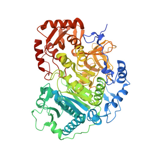
Crystal structure of Acetyl-CoA synthetase in complex with adenosine-5'-ethylphosphate from Cryptococcus neoformans var. grubii serotype A (H99)
Abendroth, J., DeBouver, N.D., Fox III, D., Esan, T.E., Hagen, T.J., Krysan, D.J., Lorimer, D.D., Horanyi, P.S., Edwards, T.E.To be published.