Structural anatomy of Protein Kinase C C1 domain interactions with diacylglycerol and other agonists.
Katti, S.S., Krieger, I.V., Ann, J., Lee, J., Sacchettini, J.C., Igumenova, T.I.(2022) Nat Commun 13: 2695-2695
- PubMed: 35577811 Search on PubMedSearch on PubMed Central
- DOI: https://doi.org/10.1038/s41467-022-30389-2
- Primary Citation Related Structures:
7KND, 7KNJ, 7KO6, 7L92, 7LCB, 7LEO, 7LF3 - PubMed Abstract:
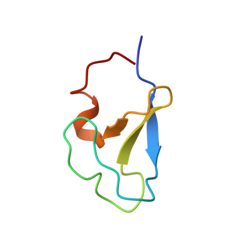
Diacylglycerol (DAG) is a versatile lipid whose 1,2-sn-stereoisomer serves both as second messenger in signal transduction pathways that control vital cellular processes, and as metabolic precursor for downstream signaling lipids such as phosphatidic acid. Effector proteins translocate to available DAG pools in the membranes by using conserved homology 1 (C1) domains as DAG-sensing modules. Yet, how C1 domains recognize and capture DAG in the complex environment of a biological membrane has remained unresolved for the 40 years since the discovery of Protein Kinase C (PKC) as the first member of the DAG effector cohort. Herein, we report the high-resolution crystal structures of a C1 domain (C1B from PKCδ) complexed to DAG and to each of four potent PKC agonists that produce different biological readouts and that command intense therapeutic interest. This structural information details the mechanisms of stereospecific recognition of DAG by the C1 domains, the functional properties of the lipid-binding site, and the identities of the key residues required for the recognition and capture of DAG and exogenous agonists. Moreover, the structures of the five C1 domain complexes provide the high-resolution guides for the design of agents that modulate the activities of DAG effector proteins.
- Department of Biochemistry and Biophysics, Texas A&M University, College Station, TX, 77840, USA.
Organizational Affiliation: