7KKW

Neutron structure of Reduced Human MnSOD


Experimental Data Snapshot

  • Method: NEUTRON DIFFRACTION
  • Resolution: 2.30 Å
  • R-Value Free: 0.302 
  • R-Value Work: 0.249 
  • R-Value Observed: 0.252 

wwPDB Validation   3D Report Full Report


This is version 2.0 of the entry. See complete history


Literature

Direct detection of coupled proton and electron transfers in human manganese superoxide dismutase.

Azadmanesh, J.Lutz, W.E.Coates, L.Weiss, K.L.Borgstahl, G.E.O.

(2021) Nat Commun 12: 2079-2079

  • DOI: https://doi.org/10.1038/s41467-021-22290-1
  • Primary Citation of Related Structures:  
    7KKS, 7KKU, 7KKW, 7KLB

  • PubMed Abstract: 

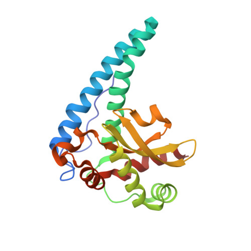
    Human manganese superoxide dismutase is a critical oxidoreductase found in the mitochondrial matrix. Concerted proton and electron transfers are used by the enzyme to rid the mitochondria of O 2 •- . The mechanisms of concerted transfer enzymes are typically unknown due to the difficulties in detecting the protonation states of specific residues and solvent molecules at particular redox states. Here, neutron diffraction of two redox-controlled manganese superoxide dismutase crystals reveal the all-atom structures of Mn 3+ and Mn 2+ enzyme forms. The structures deliver direct data on protonation changes between oxidation states of the metal. Observations include glutamine deprotonation, the involvement of tyrosine and histidine with altered pK a s, and four unusual strong-short hydrogen bonds, including a low barrier hydrogen bond. We report a concerted proton and electron transfer mechanism for human manganese superoxide dismutase from the direct visualization of active site protons in Mn 3+ and Mn 2+ redox states.


  • Organizational Affiliation

    Department of Biochemistry and Molecular Biology, 985870 Nebraska Medical Center, Omaha, NE, USA.


Macromolecules
Find similar proteins by:  (by identity cutoff)  |  3D Structure
Entity ID: 1
MoleculeChains Sequence LengthOrganismDetailsImage
Superoxide dismutase [Mn], mitochondrial
A, B
199Homo sapiensMutation(s): 0 
Gene Names: SOD2
EC: 1.15.1.1
UniProt & NIH Common Fund Data Resources
Find proteins for P04179 (Homo sapiens)
Explore P04179 
Go to UniProtKB:  P04179
PHAROS:  P04179
Entity Groups  
Sequence Clusters30% Identity50% Identity70% Identity90% Identity95% Identity100% Identity
UniProt GroupP04179
Sequence Annotations
Expand
  • Reference Sequence
Experimental Data & Validation

Experimental Data

  • Method: NEUTRON DIFFRACTION
  • Resolution: 2.30 Å
  • R-Value Free: 0.302 
  • R-Value Work: 0.249 
  • R-Value Observed: 0.252 
  • Space Group: P 61 2 2
Unit Cell:
Length ( Å )Angle ( ˚ )
a = 81.33α = 90
b = 81.33β = 90
c = 242.88γ = 120
Software Package:
Software NamePurpose
Mantiddata reduction
LaueViewdata scaling
PHENIXrefinement
PHENIXphasing

Structure Validation

View Full Validation Report


Entry History & Funding Information

Deposition Data


Funding OrganizationLocationGrant Number
National Aeronautics and Space Administration (NASA, United States)United States44-0307-1021-201
Department of Energy (DOE, United States)United States--

Revision History  (Full details and data files)

  • Version 1.0: 2021-04-21
    Type: Initial release
  • Version 1.1: 2023-10-18
    Changes: Data collection, Database references, Refinement description
  • Version 2.0: 2024-04-10
    Type: Coordinate replacement
    Reason: Model completeness
    Changes: Advisory, Atomic model, Author supporting evidence, Data collection, Derived calculations, Non-polymer description, Refinement description, Source and taxonomy, Structure summary