7KJW

Structure of HIV-1 reverse transcriptase initiation complex core with efavirenz


Experimental Data Snapshot

  • Method: ELECTRON MICROSCOPY
  • Resolution: 2.90 Å
  • Aggregation State: PARTICLE 
  • Reconstruction Method: SINGLE PARTICLE 

wwPDB Validation   3D Report Full Report


This is version 1.1 of the entry. See complete history


Literature

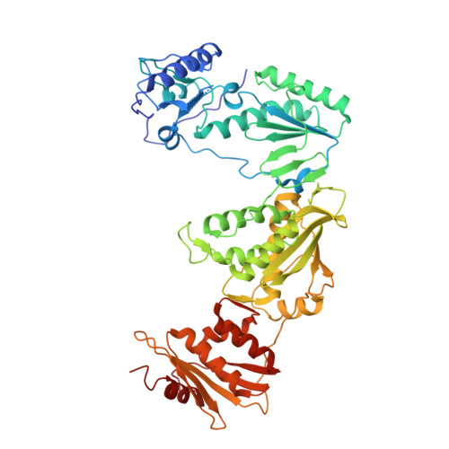
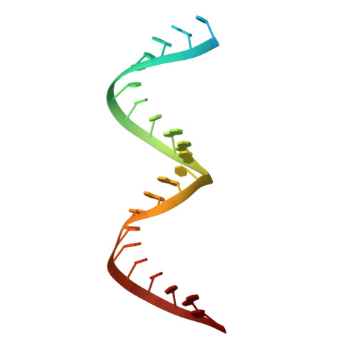
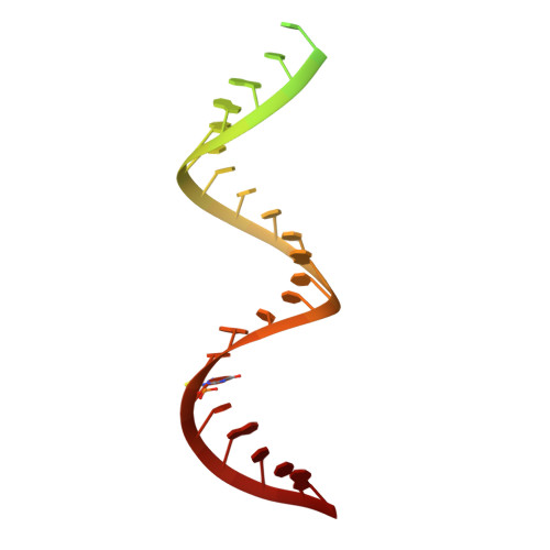
High-resolution view of HIV-1 reverse transcriptase initiation complexes and inhibition by NNRTI drugs.

Ha, B.Larsen, K.P.Zhang, J.Fu, Z.Montabana, E.Jackson, L.N.Chen, D.H.Puglisi, E.V.

(2021) Nat Commun 12: 2500-2500

  • DOI: 10.1038/s41467-021-22628-9
  • Primary Citation of Related Structures:  
    7KJV, 7KJW, 7KJX

  • PubMed Abstract: 
  • Reverse transcription of the HIV-1 viral RNA genome (vRNA) is an integral step in virus replication. Upon viral entry, HIV-1 reverse transcriptase (RT) initiates from a host tRNA Lys 3 primer bound to the vRNA genome and is the target of key antivirals, such as non-nucleoside reverse transcriptase inhibitors (NNRTIs) ...

    Reverse transcription of the HIV-1 viral RNA genome (vRNA) is an integral step in virus replication. Upon viral entry, HIV-1 reverse transcriptase (RT) initiates from a host tRNA Lys 3 primer bound to the vRNA genome and is the target of key antivirals, such as non-nucleoside reverse transcriptase inhibitors (NNRTIs). Initiation proceeds slowly with discrete pausing events along the vRNA template. Despite prior medium-resolution structural characterization of reverse transcriptase initiation complexes (RTICs), higher-resolution structures of the RTIC are needed to understand the molecular mechanisms that underlie initiation. Here we report cryo-EM structures of the core RTIC, RTIC-nevirapine, and RTIC-efavirenz complexes at 2.8, 3.1, and 2.9 Å, respectively. In combination with biochemical studies, these data suggest a basis for rapid dissociation kinetics of RT from the vRNA-tRNA Lys 3 initiation complex and reveal a specific structural mechanism of nucleic acid conformational stabilization during initiation. Finally, our results show that NNRTIs inhibit the RTIC and exacerbate discrete pausing during early reverse transcription.


    Organizational Affiliation

    Department of Structural Biology, Stanford University School of Medicine, Stanford, CA, USA. epuglisi@stanford.edu.


Macromolecules

Find similar proteins by:  (by identity cutoff)  |  3D Structure
Entity ID: 1
MoleculeChainsSequence LengthOrganismDetailsImage
Reverse transcriptase/ribonuclease HA562Human immunodeficiency virus type 1 BH10Mutation(s): 2 
Gene Names: gag-pol
EC: 2.7.7.49 (PDB Primary Data), 2.7.7.7 (PDB Primary Data), 3.1.26.13 (PDB Primary Data), 3.4.23.16 (UniProt), 3.1.13.2 (UniProt), 2.7.7 (UniProt), 3.1 (UniProt)
UniProt
Find proteins for P03366 (Human immunodeficiency virus type 1 group M subtype B (isolate BH10))
Explore P03366 
Go to UniProtKB:  P03366
Entity Groups  
Sequence Clusters30% Identity50% Identity70% Identity90% Identity95% Identity100% Identity
UniProt GroupP03366
Protein Feature View
Expand
  • Reference Sequence
Find similar proteins by:  (by identity cutoff)  |  3D Structure
Entity ID: 2
MoleculeChainsSequence LengthOrganismDetailsImage
reverse transcriptase p51 subunitB442Human immunodeficiency virus 1Mutation(s): 1 
Gene Names: gag-pol
EC: 3.4.23.16 (UniProt), 2.7.7.49 (UniProt), 2.7.7.7 (UniProt), 3.1.26.13 (UniProt), 3.1.13.2 (UniProt), 2.7.7 (UniProt), 3.1 (UniProt)
UniProt
Find proteins for P03366 (Human immunodeficiency virus type 1 group M subtype B (isolate BH10))
Explore P03366 
Go to UniProtKB:  P03366
Entity Groups  
Sequence Clusters30% Identity50% Identity70% Identity90% Identity95% Identity100% Identity
UniProt GroupP03366
Protein Feature View
Expand
  • Reference Sequence
Find similar nucleic acids by: 3D Structure
Entity ID: 3
MoleculeChainsLengthOrganismImage
HIV-1 viral RNA fragmentC26Human immunodeficiency virus 1
Protein Feature View
Expand
  • Reference Sequence
Find similar nucleic acids by: 3D Structure
Entity ID: 4
MoleculeChainsLengthOrganismImage
tRNA lysine 3 fragment with GNRA tetraloopD39Homo sapiens
Protein Feature View
Expand
  • Reference Sequence
Small Molecules
Ligands 2 Unique
IDChainsName / Formula / InChI Key2D Diagram3D Interactions
EFZ (Subject of Investigation/LOI)
Query on EFZ

Download Ideal Coordinates CCD File 
F [auth A](-)-6-CHLORO-4-CYCLOPROPYLETHYNYL-4-TRIFLUOROMETHYL-1,4-DIHYDRO-2H-3,1-BENZOXAZIN-2-ONE
C14 H9 Cl F3 N O2
XPOQHMRABVBWPR-ZDUSSCGKSA-N
 Ligand Interaction
MG
Query on MG

Download Ideal Coordinates CCD File 
E [auth A]MAGNESIUM ION
Mg
JLVVSXFLKOJNIY-UHFFFAOYSA-N
 Ligand Interaction
Experimental Data & Validation

Experimental Data

  • Method: ELECTRON MICROSCOPY
  • Resolution: 2.90 Å
  • Aggregation State: PARTICLE 
  • Reconstruction Method: SINGLE PARTICLE 
EM Software:
TaskSoftware PackageVersion
MODEL REFINEMENTPHENIX
RECONSTRUCTIONRELION

Structure Validation

View Full Validation Report


Entry History & Funding Information

Deposition Data


Funding OrganizationLocationGrant Number
National Institutes of Health/National Institute Of Allergy and Infectious Diseases (NIH/NIAID)United StatesAI150464

Revision History  (Full details and data files)

  • Version 1.0: 2021-03-17
    Type: Initial release
  • Version 1.1: 2021-05-12
    Changes: Database references