7KJO

crystal structure of PLEKHA7 PH domain biding SO4


Experimental Data Snapshot

  • Method: X-RAY DIFFRACTION
  • Resolution: 1.45 Å
  • R-Value Free: 0.171 
  • R-Value Work: 0.153 
  • R-Value Observed: 0.154 

wwPDB Validation   3D Report Full Report


This is version 1.3 of the entry. See complete history


Literature

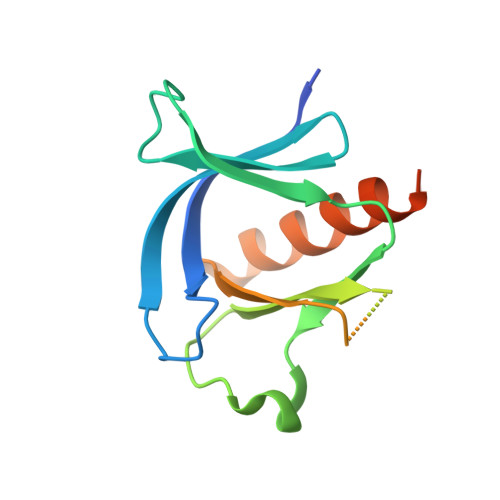
Structural basis for the association of PLEKHA7 with membrane-embedded phosphatidylinositol lipids.

Aleshin, A.E.Yao, Y.Iftikhar, A.Bobkov, A.A.Yu, J.Cadwell, G.Klein, M.G.Dong, C.Bankston, L.A.Liddington, R.C.Im, W.Powis, G.Marassi, F.M.

(2021) Structure 29: 1029

  • DOI: https://doi.org/10.1016/j.str.2021.03.018
  • Primary Citation of Related Structures:  
    7KJO, 7KJZ, 7KK7

  • PubMed Abstract: 

    PLEKHA7 (pleckstrin homology domain containing family A member 7) plays key roles in intracellular signaling, cytoskeletal organization, and cell adhesion, and is associated with multiple human cancers. The interactions of its pleckstrin homology (PH) domain with membrane phosphatidyl-inositol-phosphate (PIP) lipids are critical for proper cellular localization and function, but little is known about how PLEKHA7 and other PH domains interact with membrane-embedded PIPs. Here we describe the structural basis for recognition of membrane-bound PIPs by PLEHA7. Using X-ray crystallography, nuclear magnetic resonance, molecular dynamics simulations, and isothermal titration calorimetry, we show that the interaction of PLEKHA7 with PIPs is multivalent, distinct from a discrete one-to-one interaction, and induces PIP clustering. Our findings reveal a central role of the membrane assembly in mediating protein-PIP association and provide a roadmap for understanding how the PH domain contributes to the signaling, adhesion, and nanoclustering functions of PLEKHA7.


  • Organizational Affiliation

    Cancer Center, Sanford Burnham Prebys Medical Discovery Institute, 10901 North Torrey Pines Road, La Jolla, CA 92037, USA.


Macromolecules
Find similar proteins by:  (by identity cutoff)  |  3D Structure
Entity ID: 1
MoleculeChains Sequence LengthOrganismDetailsImage
Pleckstrin homology domain-containing family A member 7
A, B
140Homo sapiensMutation(s): 0 
Gene Names: PLEKHA7
UniProt & NIH Common Fund Data Resources
Find proteins for Q6IQ23 (Homo sapiens)
Explore Q6IQ23 
Go to UniProtKB:  Q6IQ23
PHAROS:  Q6IQ23
GTEx:  ENSG00000166689 
Entity Groups  
Sequence Clusters30% Identity50% Identity70% Identity90% Identity95% Identity100% Identity
UniProt GroupQ6IQ23
Sequence Annotations
Expand
  • Reference Sequence
Experimental Data & Validation

Experimental Data

  • Method: X-RAY DIFFRACTION
  • Resolution: 1.45 Å
  • R-Value Free: 0.171 
  • R-Value Work: 0.153 
  • R-Value Observed: 0.154 
  • Space Group: P 41
Unit Cell:
Length ( Å )Angle ( ˚ )
a = 64.83α = 90
b = 64.83β = 90
c = 59.233γ = 90
Software Package:
Software NamePurpose
PHENIXrefinement
PDB_EXTRACTdata extraction
iMOSFLMdata reduction
Aimlessdata scaling
PHASERphasing

Structure Validation

View Full Validation Report


Entry History & Funding Information

Deposition Data


Funding OrganizationLocationGrant Number
National Institutes of Health/National Cancer Institute (NIH/NCI)United StatesCA179087
National Institutes of Health/National Cancer Institute (NIH/NCI)United StatesCA160398

Revision History  (Full details and data files)

  • Version 1.0: 2021-04-07
    Type: Initial release
  • Version 1.1: 2021-05-05
    Changes: Database references
  • Version 1.2: 2021-09-15
    Changes: Database references
  • Version 1.3: 2023-10-18
    Changes: Data collection, Refinement description