


Crystal structure of GDP-mannose 4,6-dehydratase from Brucella abortus (strain 2308) in complex with Guanosine-diphosphate-rhamnose
Abendroth, J., Lorimer, D.D., Horanyi, P.S., Edwards, T.E.To be published.
Experimental Data Snapshot
Starting Model: experimental
View more details
Entity ID: 1 | |||||
|---|---|---|---|---|---|
| Molecule | Chains | Sequence Length | Organism | Details | Image |
| GDP-mannose 4,6-dehydratase | 369 | Brucella abortus 2308 | Mutation(s): 0 Gene Names: gmd, BAB1_0545 EC: 4.2.1.47 | 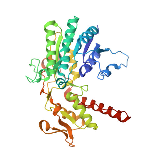 | |
UniProt | |||||
Entity Groups | |||||
| Sequence Clusters | 30% Identity50% Identity70% Identity90% Identity95% Identity100% Identity | ||||
| UniProt Group | Q2YMP3 | ||||
Sequence AnnotationsExpand | |||||
Reference Sequence | |||||
| Ligands 2 Unique | |||||
|---|---|---|---|---|---|
| ID | Chains | Name / Formula / InChI Key | 2D Diagram | 3D Interactions | |
| NAP (Subject of Investigation/LOI) Download:Ideal Coordinates CCD File | F [auth A], H [auth B], J [auth C], L [auth D] | NADP NICOTINAMIDE-ADENINE-DINUCLEOTIDE PHOSPHATE C21 H28 N7 O17 P3 XJLXINKUBYWONI-NNYOXOHSSA-N |  | ||
| GDR (Subject of Investigation/LOI) Download:Ideal Coordinates CCD File | E [auth A], G [auth B], I [auth C], K [auth D] | GUANOSINE-5'-DIPHOSPHATE-RHAMNOSE C16 H25 N5 O15 P2 LQEBEXMHBLQMDB-GDJBGNAASA-N |  | ||
| Length ( Å ) | Angle ( ˚ ) |
|---|---|
| a = 59.13 | α = 89.06 |
| b = 93.3 | β = 78.91 |
| c = 93.77 | γ = 87.51 |
| Software Name | Purpose |
|---|---|
| XDS | data reduction |
| XSCALE | data scaling |
| PHENIX | refinement |
| PDB_EXTRACT | data extraction |
| MoRDa | phasing |
| PHENIX | model building |
| Coot | model building |


