


Crystallographic structure of L,D-transpeptidase 2 from Mycobacterium tuberculosis.
Libreros, G.A., Dias, M.V.B.To be published.
Experimental Data Snapshot
Starting Model: experimental
View more details
wwPDB Validation 3D Report Full Report

Entity ID: 1 | |||||
|---|---|---|---|---|---|
| Molecule | Chains | Sequence Length | Organism | Details | Image |
| L,D-transpeptidase 2 | 408 | Mycobacterium tuberculosis H37Rv | Mutation(s): 0 Gene Names: ldtB, lppS, Rv2518c, RVBD_2518c, P425_02624 EC: 2.3.2 | 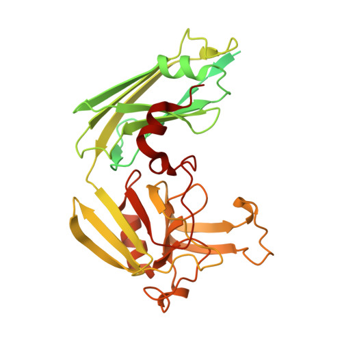 | |
UniProt | |||||
Entity Groups | |||||
| Sequence Clusters | 30% Identity50% Identity70% Identity90% Identity95% Identity100% Identity | ||||
| UniProt Group | I6Y9J2 | ||||
Sequence AnnotationsExpand | |||||
Reference Sequence | |||||
| Ligands 4 Unique | |||||
|---|---|---|---|---|---|
| ID | Chains | Name / Formula / InChI Key | 2D Diagram | 3D Interactions | |
| 0JC Download:Ideal Coordinates CCD File | C [auth A] D [auth A] E [auth A] F [auth A] J [auth B] | Di-mu-iodobis(ethylenediamine)diplatinum(II) C4 H16 I2 N4 Pt2 XXLKNYJQAYMQHB-UHFFFAOYSA-N |  | ||
| PT Download:Ideal Coordinates CCD File | I [auth B] | PLATINUM (II) ION Pt HRGDZIGMBDGFTC-UHFFFAOYSA-N |  | ||
| 6CL Download:Ideal Coordinates CCD File | H [auth B] | 6-CARBOXYLYSINE C7 H15 N2 O4 GMKMEZVLHJARHF-SYDPRGILSA-O |  | ||
| DGL Download:Ideal Coordinates CCD File | G [auth A] | D-GLUTAMIC ACID C5 H9 N O4 WHUUTDBJXJRKMK-GSVOUGTGSA-N |  | ||
| Length ( Å ) | Angle ( ˚ ) |
|---|---|
| a = 117.528 | α = 90 |
| b = 121.051 | β = 90 |
| c = 122.699 | γ = 90 |
| Software Name | Purpose |
|---|---|
| Aimless | data scaling |
| REFMAC | refinement |
| PDB_EXTRACT | data extraction |
| XDS | data reduction |
| PHASER | phasing |
| Funding Organization | Location | Grant Number |
|---|---|---|
| Fundacao para a Ciencia e a Tecnologia | Portugal | 2010/15971-3 |


