7KDX

Crystal structure of Streptomyces tokunonesis TokK with hydroxycobalamin, 5'-deoxyadenosine, and methionine


Experimental Data Snapshot

  • Method: X-RAY DIFFRACTION
  • Resolution: 1.79 Å
  • R-Value Free: 0.193 
  • R-Value Work: 0.165 
  • R-Value Observed: 0.165 

wwPDB Validation   3D Report Full Report


Ligand Structure Quality Assessment 


This is version 1.3 of the entry. See complete history


Literature

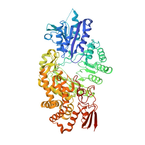
Structure of a B 12 -dependent radical SAM enzyme in carbapenem biosynthesis.

Knox, H.L.Sinner, E.K.Townsend, C.A.Boal, A.K.Booker, S.J.

(2022) Nature 602: 343-348

  • DOI: https://doi.org/10.1038/s41586-021-04392-4
  • Primary Citation of Related Structures:  
    7KDX, 7KDY

  • PubMed Abstract: 

    Carbapenems are antibiotics of last resort in the clinic. Owing to their potency and broad-spectrum activity, they are an important part of the antibiotic arsenal. The vital role of carbapenems is exemplified by the approval acquired by Merck from the US Food and Drug Administration (FDA) for the use of an imipenem combination therapy to treat the increased levels of hospital-acquired and ventilator-associated bacterial pneumonia that have occurred during the COVID-19 pandemic 1 . The C6 hydroxyethyl side chain distinguishes the clinically used carbapenems from the other classes of β-lactam antibiotics and is responsible for their low susceptibility to inactivation by occluding water from the β-lactamase active site 2 . The construction of the C6 hydroxyethyl side chain is mediated by cobalamin- or B 12 -dependent radical S-adenosylmethionine (SAM) enzymes 3 . These radical SAM methylases (RSMTs) assemble the alkyl backbone by sequential methylation reactions, and thereby underlie the therapeutic usefulness of clinically used carbapenems. Here we present X-ray crystal structures of TokK, a B 12 -dependent RSMT that catalyses three-sequential methylations during the biosynthesis of asparenomycin A. These structures, which contain the two metallocofactors of the enzyme and were determined in the presence and absence of a carbapenam substrate, provide a visualization of a B 12 -dependent RSMT that uses the radical mechanism that is shared by most of these enzymes. The structures provide insight into the stereochemistry of initial C6 methylation and suggest that substrate positioning governs the rate of each methylation event.


  • Organizational Affiliation

    Department of Chemistry, Pennsylvania State University, University Park, PA, USA.


Macromolecules
Find similar proteins by:  (by identity cutoff)  |  3D Structure
Entity ID: 1
MoleculeChains Sequence LengthOrganismDetailsImage
Methyltransferase TokK
A, B
681Streptomyces tokunonensisMutation(s): 0 
Gene Names: tokK
UniProt
Find proteins for A0A6B9HEI0 (Streptomyces tokunonensis)
Explore A0A6B9HEI0 
Go to UniProtKB:  A0A6B9HEI0
Entity Groups  
Sequence Clusters30% Identity50% Identity70% Identity90% Identity95% Identity100% Identity
UniProt GroupA0A6B9HEI0
Sequence Annotations
Expand
  • Reference Sequence
Small Molecules
Ligands 7 Unique
IDChains Name / Formula / InChI Key2D Diagram3D Interactions
B12 (Subject of Investigation/LOI)
Query on B12

Download Ideal Coordinates CCD File 
E [auth A],
P [auth B]
COBALAMIN
C62 H89 Co N13 O14 P
LKVIQTCSMMVGFU-DWSMJLPVSA-N
SF4 (Subject of Investigation/LOI)
Query on SF4

Download Ideal Coordinates CCD File 
C [auth A],
Q [auth B]
IRON/SULFUR CLUSTER
Fe4 S4
LJBDFODJNLIPKO-UHFFFAOYSA-N
5AD (Subject of Investigation/LOI)
Query on 5AD

Download Ideal Coordinates CCD File 
D [auth A],
R [auth B]
5'-DEOXYADENOSINE
C10 H13 N5 O3
XGYIMTFOTBMPFP-KQYNXXCUSA-N
MET (Subject of Investigation/LOI)
Query on MET

Download Ideal Coordinates CCD File 
F [auth A],
S [auth B]
METHIONINE
C5 H11 N O2 S
FFEARJCKVFRZRR-BYPYZUCNSA-N
EDO
Query on EDO

Download Ideal Coordinates CCD File 
AA [auth B]
BA [auth B]
CA [auth B]
DA [auth B]
EA [auth B]
AA [auth B],
BA [auth B],
CA [auth B],
DA [auth B],
EA [auth B],
FA [auth B],
G [auth A],
H [auth A],
I [auth A],
J [auth A],
K [auth A],
L [auth A],
M [auth A],
O [auth B],
T [auth B],
U [auth B],
V [auth B],
W [auth B],
X [auth B],
Y [auth B],
Z [auth B]
1,2-ETHANEDIOL
C2 H6 O2
LYCAIKOWRPUZTN-UHFFFAOYSA-N
K
Query on K

Download Ideal Coordinates CCD File 
IA [auth B],
N [auth A]
POTASSIUM ION
K
NPYPAHLBTDXSSS-UHFFFAOYSA-N
CL
Query on CL

Download Ideal Coordinates CCD File 
GA [auth B],
HA [auth B]
CHLORIDE ION
Cl
VEXZGXHMUGYJMC-UHFFFAOYSA-M
Experimental Data & Validation

Experimental Data

  • Method: X-RAY DIFFRACTION
  • Resolution: 1.79 Å
  • R-Value Free: 0.193 
  • R-Value Work: 0.165 
  • R-Value Observed: 0.165 
  • Space Group: P 1 21 1
Unit Cell:
Length ( Å )Angle ( ˚ )
a = 68.644α = 90
b = 137.739β = 90.47
c = 77.914γ = 90
Software Package:
Software NamePurpose
PHENIXrefinement
PDB_EXTRACTdata extraction
HKL-2000data scaling
AutoSolphasing
HKL-2000data reduction

Structure Validation

View Full Validation Report


Ligand Structure Quality Assessment 


Entry History & Funding Information

Deposition Data


Funding OrganizationLocationGrant Number
National Institutes of Health/National Institute of General Medical Sciences (NIH/NIGMS)United StatesGM-12259

Revision History  (Full details and data files)

  • Version 1.0: 2022-01-12
    Type: Initial release
  • Version 1.1: 2022-02-02
    Changes: Database references
  • Version 1.2: 2022-02-16
    Changes: Database references
  • Version 1.3: 2022-02-23
    Changes: Database references