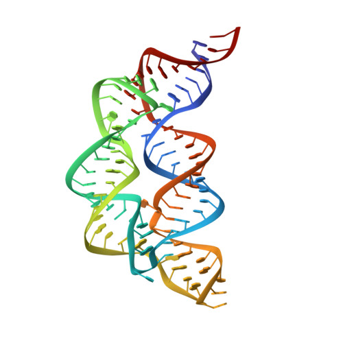
Tying the knot in the tetrahydrofolate (THF) riboswitch: A molecular basis for gene regulation.
Wilt, H.M., Yu, P., Tan, K., Wang, Y.X., Stagno, J.R.(2021) J Struct Biol 213: 107703-107703
- PubMed: 33571639 Search on PubMedSearch on PubMed Central
- DOI: https://doi.org/10.1016/j.jsb.2021.107703
- Primary Citation Related Structures:
7KD1 - PubMed Abstract:
Effective gene regulation by the tetrahydrofolate riboswitch depends not only on ligand affinity but also on the kinetics of ligand association, which involves two cooperative binding sites. We have determined a 1.9-Å resolution crystal structure of the ligand-free THF riboswitch aptamer. The pseudoknot binding site 'unwinds' in the absence of ligand, whereby the adjacent helical domains (P1, P2, and P3) become disjointed, resulting in rotation and misalignment of the gene-regulatory P1 helix with respect to P3. In contrast, the second binding site at the three-way junction, which is the first to fold, is structurally conserved between apo and holo forms. This suggests a kinetic role for this site, in which binding of the first ligand molecule to the stably folded three-way junction promotes formation of the regulatory pseudoknot site and subsequent binding of the second molecule. As such, these findings provide a molecular basis for both conformational switching and kinetic control.
- Structural Biophysics Laboratory, Center for Cancer Research, National Cancer Institute, Frederick, MD 21702, USA.
Organizational Affiliation: