7KAW

Crystal structure of OhyA-PEG400-FAD complex from Staphylococcus aureus

  • Classification: OXIDOREDUCTASE
  • Organism(s): Staphylococcus aureus
  • Expression System: Escherichia coli
  • Mutation(s): No 

  • Deposited: 2020-10-01 Released: 2021-01-06 
  • Deposition Author(s): Radka, C.D., Rock, C.O.
  • Funding Organization(s): National Institutes of Health/National Institute of General Medical Sciences (NIH/NIGMS), National Institutes of Health/National Cancer Institute (NIH/NCI)

Experimental Data Snapshot

  • Method: X-RAY DIFFRACTION
  • Resolution: 2.10 Å
  • R-Value Free: 0.232 
  • R-Value Work: 0.189 
  • R-Value Observed: 0.190 

wwPDB Validation   3D Report Full Report


Ligand Structure Quality Assessment 


This is version 1.3 of the entry. See complete history


Literature

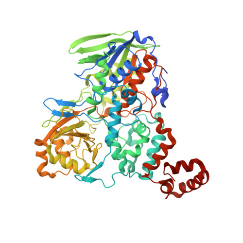
Structure and mechanism of Staphylococcus aureus oleate hydratase (OhyA).

Radka, C.D.Batte, J.L.Frank, M.W.Young, B.M.Rock, C.O.

(2021) J Biol Chem 296: 100252-100252

  • DOI: https://doi.org/10.1074/jbc.RA120.016818
  • Primary Citation of Related Structures:  
    7KAV, 7KAW, 7KAX, 7KAY, 7KAZ

  • PubMed Abstract: 

    Flavin adenine dinucleotide (FAD)-dependent bacterial oleate hydratases (OhyAs) catalyze the addition of water to isolated fatty acid carbon-carbon double bonds. Staphylococcus aureus uses OhyA to counteract the host innate immune response by inactivating antimicrobial unsaturated fatty acids. Mechanistic information explaining how OhyAs catalyze regiospecific and stereospecific hydration is required to understand their biological functions and the potential for engineering new products. In this study, we deduced the catalytic mechanism of OhyA from multiple structures of S. aureus OhyA in binary and ternary complexes with combinations of ligands along with biochemical analyses of relevant mutants. The substrate-free state shows Arg81 is the gatekeeper that controls fatty acid entrance to the active site. FAD binding engages the catalytic loop to simultaneously rotate Glu82 into its active conformation and Arg81 out of the hydrophobic substrate tunnel, allowing the fatty acid to rotate into the active site. FAD binding also dehydrates the active site, leaving a single water molecule connected to Glu82. This active site water is a hydronium ion based on the analysis of its hydrogen bond network in the OhyA•PEG400•FAD complex. We conclude that OhyA accelerates acid-catalyzed alkene hydration by positioning the fatty acid double bond to attack the active site hydronium ion, followed by the addition of water to the transient carbocation intermediate. Structural transitions within S. aureus OhyA channel oleate to the active site, curl oleate around the substrate water, and stabilize the hydroxylated product to inactivate antimicrobial fatty acids.


  • Organizational Affiliation

    The Department of Infectious Diseases, St Jude Children's Research Hospital, Memphis, Tennessee, USA.


Macromolecules
Find similar proteins by:  (by identity cutoff)  |  3D Structure
Entity ID: 1
MoleculeChains Sequence LengthOrganismDetailsImage
Oleate hydrataseA [auth C],
B,
C [auth A]
611Staphylococcus aureusMutation(s): 0 
Gene Names: 
UniProt
Find proteins for A0A0D6GJV1 (Staphylococcus aureus)
Explore A0A0D6GJV1 
Go to UniProtKB:  A0A0D6GJV1
Entity Groups  
Sequence Clusters30% Identity50% Identity70% Identity90% Identity95% Identity100% Identity
UniProt GroupA0A0D6GJV1
Sequence Annotations
Expand
  • Reference Sequence
Small Molecules
Ligands 3 Unique
IDChains Name / Formula / InChI Key2D Diagram3D Interactions
FAD (Subject of Investigation/LOI)
Query on FAD

Download Ideal Coordinates CCD File 
E [auth C],
H [auth B]
FLAVIN-ADENINE DINUCLEOTIDE
C27 H33 N9 O15 P2
VWWQXMAJTJZDQX-UYBVJOGSSA-N
P6G (Subject of Investigation/LOI)
Query on P6G

Download Ideal Coordinates CCD File 
D [auth C],
G [auth B],
J [auth A]
HEXAETHYLENE GLYCOL
C12 H26 O7
IIRDTKBZINWQAW-UHFFFAOYSA-N
GOL
Query on GOL

Download Ideal Coordinates CCD File 
F [auth C],
I [auth B],
K [auth A],
L [auth A]
GLYCEROL
C3 H8 O3
PEDCQBHIVMGVHV-UHFFFAOYSA-N
Experimental Data & Validation

Experimental Data

  • Method: X-RAY DIFFRACTION
  • Resolution: 2.10 Å
  • R-Value Free: 0.232 
  • R-Value Work: 0.189 
  • R-Value Observed: 0.190 
  • Space Group: C 1 2 1
Unit Cell:
Length ( Å )Angle ( ˚ )
a = 188.08α = 90
b = 112.919β = 116.97
c = 119.059γ = 90
Software Package:
Software NamePurpose
PHENIXrefinement
PDB_EXTRACTdata extraction
KYLINdata reduction
KYLINdata scaling
PHASERphasing

Structure Validation

View Full Validation Report


Ligand Structure Quality Assessment 


Entry History & Funding Information

Deposition Data


Funding OrganizationLocationGrant Number
National Institutes of Health/National Institute of General Medical Sciences (NIH/NIGMS)United StatesGM034496
National Institutes of Health/National Cancer Institute (NIH/NCI)United StatesCA21765

Revision History  (Full details and data files)

  • Version 1.0: 2021-01-06
    Type: Initial release
  • Version 1.1: 2021-01-13
    Changes: Database references
  • Version 1.2: 2021-04-28
    Changes: Database references
  • Version 1.3: 2023-10-18
    Changes: Data collection, Database references, Refinement description