


Crystal Structure of Dihydrofolate reductase from Mycobacterium kansasii in complex with NADP and inhibitor P218
Abendroth, J., Dranow, D.M., Santhakumar, V., Walpole, C., Lorimer, D.D., Horanyi, P.S., Edwards, T.E.To be published.
Experimental Data Snapshot
Starting Model: experimental
View more details
Entity ID: 1 | |||||
|---|---|---|---|---|---|
| Molecule | Chains | Sequence Length | Organism | Details | Image |
| Dihydrofolate reductase | 167 | Mycobacterium kansasii | Mutation(s): 0 Gene Names: folA, BZL29_1474, BZL30_0350 EC: 1.5.1.3 | 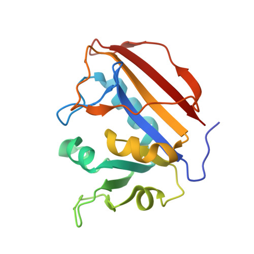 | |
UniProt | |||||
Entity Groups | |||||
| Sequence Clusters | 30% Identity50% Identity70% Identity90% Identity95% Identity100% Identity | ||||
| UniProt Group | A0A653EQ34 | ||||
Sequence AnnotationsExpand | |||||
Reference Sequence | |||||
| Ligands 3 Unique | |||||
|---|---|---|---|---|---|
| ID | Chains | Name / Formula / InChI Key | 2D Diagram | 3D Interactions | |
| NAP (Subject of Investigation/LOI) Download:Ideal Coordinates CCD File | C [auth A], F [auth B] | NADP NICOTINAMIDE-ADENINE-DINUCLEOTIDE PHOSPHATE C21 H28 N7 O17 P3 XJLXINKUBYWONI-NNYOXOHSSA-N |  | ||
| MMV (Subject of Investigation/LOI) Download:Ideal Coordinates CCD File | D [auth A], G [auth B] | 3-(2-{3-[(2,4-diamino-6-ethylpyrimidin-5-yl)oxy]propoxy}phenyl)propanoic acid C18 H24 N4 O4 VDGXZSSDCDPCRF-UHFFFAOYSA-N |  | ||
| EDO Download:Ideal Coordinates CCD File | E [auth A], H [auth B] | 1,2-ETHANEDIOL C2 H6 O2 LYCAIKOWRPUZTN-UHFFFAOYSA-N |  | ||
| Length ( Å ) | Angle ( ˚ ) |
|---|---|
| a = 62.1 | α = 90 |
| b = 66.25 | β = 90 |
| c = 86.12 | γ = 90 |
| Software Name | Purpose |
|---|---|
| XDS | data reduction |
| XSCALE | data scaling |
| PHENIX | refinement |
| PDB_EXTRACT | data extraction |
| MoRDa | phasing |
| PHENIX | model building |
| Coot | model building |


