7K5Q

Bst DNA polymerase I time-resolved structure, 8 min post dATP addition


Experimental Data Snapshot

  • Method: X-RAY DIFFRACTION
  • Resolution: 2.00 Å
  • R-Value Free: 0.246 
  • R-Value Work: 0.194 
  • R-Value Observed: 0.195 

wwPDB Validation   3D Report Full Report


Ligand Structure Quality Assessment 


This is version 1.2 of the entry. See complete history


Literature

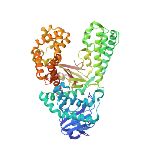
Following replicative DNA synthesis by time-resolved X-ray crystallography.

Chim, N.Meza, R.A.Trinh, A.M.Yang, K.Chaput, J.C.

(2021) Nat Commun 12: 2641-2641

  • DOI: https://doi.org/10.1038/s41467-021-22937-z
  • Primary Citation of Related Structures:  
    7K5O, 7K5P, 7K5Q, 7K5R, 7K5S, 7K5T, 7K5U

  • PubMed Abstract: 

    The mechanism of DNA synthesis has been inferred from static structures, but the absence of temporal information raises longstanding questions about the order of events in one of life's most central processes. Here we follow the reaction pathway of a replicative DNA polymerase using time-resolved X-ray crystallography to elucidate the order and transition between intermediates. In contrast to the canonical model, the structural changes observed in the time-lapsed images reveal a catalytic cycle in which translocation precedes catalysis. The translocation step appears to follow a push-pull mechanism where the O-O1 loop of the finger subdomain acts as a pawl to facilitate unidirectional movement along the template with conserved tyrosine residues 714 and 719 functioning as tandem gatekeepers of DNA synthesis. The structures capture the precise order of critical events that may be a general feature of enzymatic catalysis among replicative DNA polymerases.


  • Organizational Affiliation

    Department of Pharmaceutical Sciences, University of California, Irvine, CA, USA.


Macromolecules

Find similar proteins by:  (by identity cutoff)  |  3D Structure
Entity ID: 3
MoleculeChains Sequence LengthOrganismDetailsImage
DNA polymerase IC [auth A]580Geobacillus stearothermophilusMutation(s): 1 
Gene Names: polA
EC: 2.7.7.7
UniProt
Find proteins for E1C9K5 (Geobacillus stearothermophilus)
Explore E1C9K5 
Go to UniProtKB:  E1C9K5
Entity Groups  
Sequence Clusters30% Identity50% Identity70% Identity90% Identity95% Identity100% Identity
UniProt GroupE1C9K5
Sequence Annotations
Expand
  • Reference Sequence

Find similar nucleic acids by:  Sequence   |   3D Structure  

Entity ID: 1
MoleculeChains LengthOrganismImage
DNA (5'-D(P*G*CP*GP*AP*TP*CP*AP*CP*GP*T)-3')A [auth P]10synthetic construct
Sequence Annotations
Expand
  • Reference Sequence

Find similar nucleic acids by:  Sequence   |   3D Structure  

Entity ID: 2
MoleculeChains LengthOrganismImage
DNA (5'-D(P*GP*TP*AP*CP*GP*TP*GP*AP*TP*CP*GP*CP*A)-3')B [auth T]16synthetic construct
Sequence Annotations
Expand
  • Reference Sequence
Experimental Data & Validation

Experimental Data

  • Method: X-RAY DIFFRACTION
  • Resolution: 2.00 Å
  • R-Value Free: 0.246 
  • R-Value Work: 0.194 
  • R-Value Observed: 0.195 
  • Space Group: P 21 21 21
Unit Cell:
Length ( Å )Angle ( ˚ )
a = 88.2α = 90
b = 93.988β = 90
c = 105.724γ = 90
Software Package:
Software NamePurpose
PHENIXrefinement
autoXDSdata reduction
autoXDSdata scaling
PHASERphasing

Structure Validation

View Full Validation Report


Ligand Structure Quality Assessment 


Entry History & Funding Information

Deposition Data


Funding OrganizationLocationGrant Number
National Science Foundation (NSF, United States)United StatesCHE 2001434

Revision History  (Full details and data files)

  • Version 1.0: 2021-03-03
    Type: Initial release
  • Version 1.1: 2021-05-19
    Changes: Database references
  • Version 1.2: 2023-10-18
    Changes: Data collection, Database references, Refinement description