7K4M

Crystal structure of MetAP2 Modified Hemoglobin S


Experimental Data Snapshot

  • Method: X-RAY DIFFRACTION
  • Resolution: 2.50 Å
  • R-Value Free: 0.323 
  • R-Value Work: 0.239 
  • R-Value Observed: 0.243 

wwPDB Validation   3D Report Full Report


Ligand Structure Quality Assessment 


This is version 1.2 of the entry. See complete history


Literature

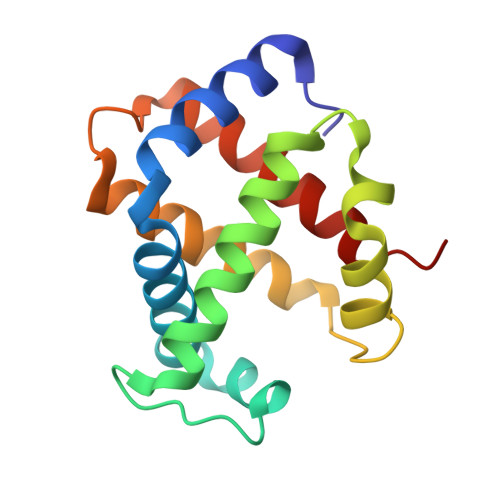
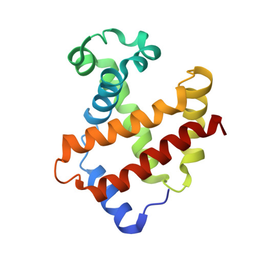
MetAP2 inhibition modifies hemoglobin S to delay polymerization and improves blood flow in sickle cell disease.

Demers, M.Sturtevant, S.Guertin, K.R.Gupta, D.Desai, K.Vieira, B.F.Li, W.Hicks, A.Ismail, A.Goncalves, B.P.Di Caprio, G.Schonbrun, E.Hansen, S.Musayev, F.N.Safo, M.K.Wood, D.K.Higgins, J.M.Light, D.R.

(2021) Blood Adv 5: 1388-1402

  • DOI: https://doi.org/10.1182/bloodadvances.2020003670
  • Primary Citation of Related Structures:  
    7K4M

  • PubMed Abstract: 

    Sickle cell disease (SCD) is associated with hemolysis, vascular inflammation, and organ damage. Affected patients experience chronic painful vaso-occlusive events requiring hospitalization. Hypoxia-induced polymerization of sickle hemoglobin S (HbS) contributes to sickling of red blood cells (RBCs) and disease pathophysiology. Dilution of HbS with nonsickling hemoglobin or hemoglobin with increased oxygen affinity, such as fetal hemoglobin or HbS bound to aromatic aldehydes, is clinically beneficial in decreasing polymerization. We investigated a novel alternate approach to modify HbS and decrease polymerization by inhibiting methionine aminopeptidase 2 (MetAP2), which cleaves the initiator methionine (iMet) from Val1 of α-globin and βS-globin. Kinetic studies with MetAP2 show that βS-globin is a fivefold better substrate than α-globin. Knockdown of MetAP2 in human umbilical cord blood-derived erythroid progenitor 2 cells shows more extensive modification of α-globin than β-globin, consistent with kinetic data. Treatment of human erythroid cells in vitro or Townes SCD mice in vivo with selective MetAP2 inhibitors extensively modifies both globins with N-terminal iMet and acetylated iMet. HbS modification by MetAP2 inhibition increases oxygen affinity, as measured by decreased oxygen tension at which hemoglobin is 50% saturated. Acetyl-iMet modification on βS-globin delays HbS polymerization under hypoxia. MetAP2 inhibitor-treated Townes mice reach 50% total HbS modification, significantly increasing the affinity of RBCs for oxygen, increasing whole blood single-cell RBC oxygen saturation, and decreasing fractional flow velocity losses in blood rheology under decreased oxygen pressures. Crystal structures of modified HbS variants show stabilization of the nonpolymerizing high O2-affinity R2 state, explaining modified HbS antisickling activity. Further study of MetAP2 inhibition as a potential therapeutic target for SCD is warranted.


  • Organizational Affiliation

    Department of Rare Blood Disorders, Sanofi, Waltham, MA.


Macromolecules
Find similar proteins by:  (by identity cutoff)  |  3D Structure
Entity ID: 1
MoleculeChains Sequence LengthOrganismDetailsImage
Hemoglobin subunit alpha
A, C, E, G, I
142Homo sapiensMutation(s): 0 
UniProt & NIH Common Fund Data Resources
Find proteins for P69905 (Homo sapiens)
Explore P69905 
Go to UniProtKB:  P69905
PHAROS:  P69905
Entity Groups  
Sequence Clusters30% Identity50% Identity70% Identity90% Identity95% Identity100% Identity
UniProt GroupP69905
Sequence Annotations
Expand
  • Reference Sequence
Find similar proteins by:  (by identity cutoff)  |  3D Structure
Entity ID: 2
MoleculeChains Sequence LengthOrganismDetailsImage
Hemoglobin subunit beta
B, D, F, H, J
148Homo sapiensMutation(s): 1 
UniProt & NIH Common Fund Data Resources
Find proteins for P68871 (Homo sapiens)
Explore P68871 
Go to UniProtKB:  P68871
PHAROS:  P68871
GTEx:  ENSG00000244734 
Entity Groups  
Sequence Clusters30% Identity50% Identity70% Identity90% Identity95% Identity100% Identity
UniProt GroupP68871
Sequence Annotations
Expand
  • Reference Sequence
Small Molecules
Ligands 2 Unique
IDChains Name / Formula / InChI Key2D Diagram3D Interactions
HEM (Subject of Investigation/LOI)
Query on HEM

Download Ideal Coordinates CCD File 
AA [auth I]
CA [auth J]
L [auth A]
N [auth B]
P [auth C]
AA [auth I],
CA [auth J],
L [auth A],
N [auth B],
P [auth C],
R [auth D],
S [auth E],
U [auth F],
W [auth G],
Y [auth H]
PROTOPORPHYRIN IX CONTAINING FE
C34 H32 Fe N4 O4
KABFMIBPWCXCRK-RGGAHWMASA-L
CMO (Subject of Investigation/LOI)
Query on CMO

Download Ideal Coordinates CCD File 
BA [auth J]
K [auth A]
M [auth B]
O [auth C]
Q [auth D]
BA [auth J],
K [auth A],
M [auth B],
O [auth C],
Q [auth D],
T [auth F],
V [auth G],
X [auth H],
Z [auth I]
CARBON MONOXIDE
C O
UGFAIRIUMAVXCW-UHFFFAOYSA-N
Experimental Data & Validation

Experimental Data

  • Method: X-RAY DIFFRACTION
  • Resolution: 2.50 Å
  • R-Value Free: 0.323 
  • R-Value Work: 0.239 
  • R-Value Observed: 0.243 
  • Space Group: I 1 2 1
Unit Cell:
Length ( Å )Angle ( ˚ )
a = 134.844α = 90
b = 53.846β = 91.73
c = 195.64γ = 90
Software Package:
Software NamePurpose
CrysalisProdata collection
Aimlessdata scaling
PHASERphasing
PHENIXrefinement
PDB_EXTRACTdata extraction

Structure Validation

View Full Validation Report


Ligand Structure Quality Assessment 


Entry History & Funding Information

Deposition Data


Funding OrganizationLocationGrant Number
National Institutes of Health/National Institute on Alcohol Abuse and Alcoholism (NIH/NIAAA)United StatesS10OD021756

Revision History  (Full details and data files)

  • Version 1.0: 2021-10-13
    Type: Initial release
  • Version 1.1: 2022-04-27
    Changes: Database references
  • Version 1.2: 2023-10-18
    Changes: Data collection, Refinement description