7K1K | pdb_00007k1k

CryoEM structure of inactivated-form DNA-PK (Complex IV)


Experimental Data Snapshot

  • Method: ELECTRON MICROSCOPY
  • Resolution: 4.10 Å
  • Aggregation State: PARTICLE 
  • Reconstruction Method: SINGLE PARTICLE 

wwPDB Validation 3D Report Full Report

Validation slider image for 7K1K

This is version 1.4 of the entry. See complete history

Literature

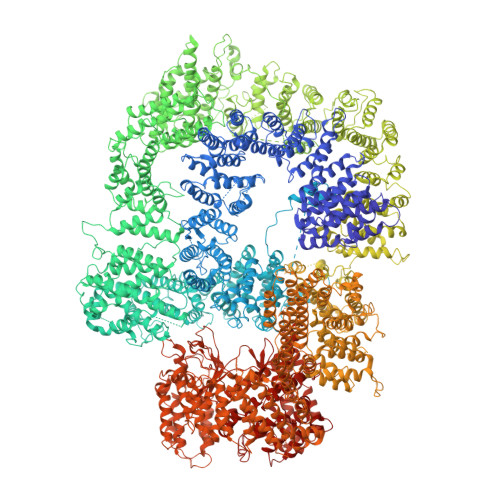
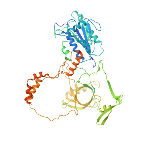
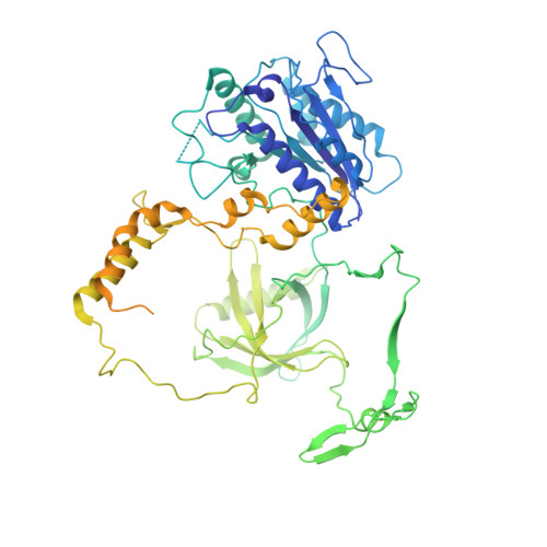
Structure of an activated DNA-PK and its implications for NHEJ.

Chen, X.Xu, X.Chen, Y.Cheung, J.C.Wang, H.Jiang, J.de Val, N.Fox, T.Gellert, M.Yang, W.

(2021) Mol Cell 81: 801-810.e3

  • DOI: https://doi.org/10.1016/j.molcel.2020.12.015
  • Primary Citation Related Structures: 
    7K0Y, 7K10, 7K11, 7K17, 7K19, 7K1B, 7K1J, 7K1K, 7K1N

  • PubMed Abstract: 

    DNA-dependent protein kinase (DNA-PK), like all phosphatidylinositol 3-kinase-related kinases (PIKKs), is composed of conserved FAT and kinase domains (FATKINs) along with solenoid structures made of HEAT repeats. These kinases are activated in response to cellular stress signals, but the mechanisms governing activation and regulation remain unresolved. For DNA-PK, all existing structures represent inactive states with resolution limited to 4.3 Å at best. Here, we report the cryoelectron microscopy (cryo-EM) structures of DNA-PKcs (DNA-PK catalytic subunit) bound to a DNA end or complexed with Ku70/80 and DNA in both inactive and activated forms at resolutions of 3.7 Å overall and 3.2 Å for FATKINs. These structures reveal the sequential transition of DNA-PK from inactive to activated forms. Most notably, activation of the kinase involves previously unknown stretching and twisting within individual solenoid segments and loosens DNA-end binding. This unprecedented structural plasticity of helical repeats may be a general regulatory mechanism of HEAT-repeat proteins.


  • Organizational Affiliation
    • Laboratory of Molecular Biology, NIDDK, National Institutes of Health, Bethesda, MD 20892, USA.

Macromolecule Content 

  • Total Structure Weight: 646.97 kDa 
  • Atom Count: 38,243 
  • Modeled Residue Count: 4,694 
  • Deposited Residue Count: 5,549 
  • Unique protein chains: 3
  • Unique nucleic acid chains: 2

Macromolecules


Find similar proteins by:|  3D Structure
Entity ID: 1
MoleculeChains  Sequence LengthOrganismDetailsImage
DNA-dependent protein kinase catalytic subunit4,128Homo sapiensMutation(s): 0 
EC: 2.7.11.1
UniProt & NIH Common Fund Data Resources
Find proteins for P78527 (Homo sapiens)
Explore P78527 
Go to UniProtKB:  P78527
PHAROS:  P78527
GTEx:  ENSG00000253729 
Entity Groups
Sequence Clusters30% Identity50% Identity70% Identity90% Identity95% Identity100% Identity
UniProt GroupP78527
Sequence Annotations
Expand
Reference Sequence
Find similar proteins by:|  3D Structure
Entity ID: 2
MoleculeChains  Sequence LengthOrganismDetailsImage
X-ray repair cross-complementing protein 6609Homo sapiensMutation(s): 0 
Gene Names: XRCC6G22P1
EC: 3.6.4 (PDB Primary Data), 4.2.99 (PDB Primary Data), 5.6.2.4 (UniProt), 4.2.99.18 (UniProt)
UniProt & NIH Common Fund Data Resources
Find proteins for P12956 (Homo sapiens)
Explore P12956 
Go to UniProtKB:  P12956
PHAROS:  P12956
GTEx:  ENSG00000196419 
Entity Groups
Sequence Clusters30% Identity50% Identity70% Identity90% Identity95% Identity100% Identity
UniProt GroupP12956
Sequence Annotations
Expand
Reference Sequence
Find similar proteins by:|  3D Structure
Entity ID: 3
MoleculeChains  Sequence LengthOrganismDetailsImage
X-ray repair cross-complementing protein 5732Homo sapiensMutation(s): 0 
Gene Names: XRCC5G22P2
EC: 3.6.4 (PDB Primary Data), 5.6.2.4 (UniProt), 4.2.99.18 (UniProt)
UniProt & NIH Common Fund Data Resources
Find proteins for P13010 (Homo sapiens)
Explore P13010 
Go to UniProtKB:  P13010
PHAROS:  P13010
GTEx:  ENSG00000079246 
Entity Groups
Sequence Clusters30% Identity50% Identity70% Identity90% Identity95% Identity100% Identity
UniProt GroupP13010
Sequence Annotations
Expand
Reference Sequence
Find similar nucleic acids by:  Sequence
Entity ID: 4
MoleculeChains LengthOrganismImage
DNA (5'-D(*GP*CP*AP*TP*GP*CP*TP*CP*TP*AP*CP*TP*GP*CP*TP*TP*CP*GP*AP*TP*AP*TP*CP*G)-3')
D, F
24synthetic construct
Sequence Annotations
Expand
Reference Sequence
Find similar nucleic acids by:  Sequence
Entity ID: 5
MoleculeChains LengthOrganismImage
DNA (5'-D(*AP*AP*GP*CP*AP*GP*TP*AP*GP*AP*GP*CP*A)-3')
E, G
16synthetic construct
Sequence Annotations
Expand
Reference Sequence

Experimental Data & Validation

Experimental Data

  • Method: ELECTRON MICROSCOPY
  • Resolution: 4.10 Å
  • Aggregation State: PARTICLE 
  • Reconstruction Method: SINGLE PARTICLE 
EM Software:
TaskSoftware PackageVersion
MODEL REFINEMENTPHENIX

Structure Validation

View Full Validation Report


Entry History 

& Funding Information

Deposition Data


Funding OrganizationLocationGrant Number
National Institutes of Health/National Institute of Diabetes and Digestive and Kidney Disease (NIH/NIDDK)United States--

Revision History  (Full details and data files)

  • Version 1.0: 2021-01-06
    Type: Initial release
  • Version 1.1: 2021-01-13
    Changes: Database references
  • Version 1.2: 2021-03-03
    Changes: Database references
  • Version 1.3: 2024-03-06
    Changes: Data collection, Database references
  • Version 1.4: 2025-05-28
    Changes: Data collection, Structure summary