7K15 | pdb_00007k15

Crystal structure of the Human Leukotriene B4 Receptor 1 in Complex with Selective Antagonist MK-D-046


Experimental Data Snapshot

  • Method: X-RAY DIFFRACTION
  • Resolution: 2.88 Å
  • R-Value Free: 
    0.261 (Depositor), 0.266 (DCC) 
  • R-Value Work: 
    0.212 (Depositor), 0.216 (DCC) 
  • R-Value Observed: 
    0.214 (Depositor) 

Starting Models: experimental
View more details

wwPDB Validation 3D Report Full Report

Validation slider image for 7K15

Ligand Structure Quality Assessment 


This is version 1.3 of the entry. See complete history

Literature

Structural insights on ligand recognition at the human leukotriene B4 receptor 1.

Michaelian, N.Sadybekov, A.Besserer-Offroy, E.Han, G.W.Krishnamurthy, H.Zamlynny, B.A.Fradera, X.Siliphaivanh, P.Presland, J.Spencer, K.B.Soisson, S.M.Popov, P.Sarret, P.Katritch, V.Cherezov, V.

(2021) Nat Commun 12: 2971-2971

  • DOI: https://doi.org/10.1038/s41467-021-23149-1
  • Primary Citation Related Structures: 
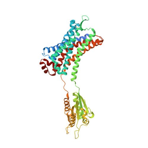
    7K15

  • PubMed Abstract: 

    The leukotriene B4 receptor 1 (BLT1) regulates the recruitment and chemotaxis of different cell types and plays a role in the pathophysiology of infectious, allergic, metabolic, and tumorigenic human diseases. Here we present a crystal structure of human BLT1 (hBLT1) in complex with a selective antagonist MK-D-046, developed for the treatment of type 2 diabetes and other inflammatory conditions. Comprehensive analysis of the structure and structure-activity relationship data, reinforced by site-directed mutagenesis and docking studies, reveals molecular determinants of ligand binding and selectivity toward different BLT receptor subtypes and across species. The structure helps to identify a putative membrane-buried ligand access channel as well as potential receptor binding modes of endogenous agonists. These structural insights of hBLT1 enrich our understanding of its ligand recognition and open up future avenues in structure-based drug design.


  • Organizational Affiliation
    • Bridge Institute, USC Michelson Center for Convergent Bioscience, University of Southern California, Los Angeles, CA, USA.

Macromolecule Content 

  • Total Structure Weight: 56.14 kDa 
  • Atom Count: 3,624 
  • Modeled Residue Count: 455 
  • Deposited Residue Count: 490 
  • Unique protein chains: 1

Macromolecules

Find similar proteins by:|  3D Structure
Entity ID: 1
MoleculeChains  Sequence LengthOrganismDetailsImage
Leukotriene B4 receptor 1,Flavodoxin,Leukotriene B4 receptor 1490Homo sapiensNitratidesulfovibrio vulgaris str. HildenboroughMutation(s): 7 
Gene Names: LTB4RBLTBLT1BLTRCMKRL1GPR16P2RY7DVU_2680
Membrane Entity: Yes 
UniProt & NIH Common Fund Data Resources
Find proteins for P00323 (Nitratidesulfovibrio vulgaris (strain ATCC 29579 / DSM 644 / CCUG 34227 / NCIMB 8303 / VKM B-1760 / Hildenborough))
Explore P00323 
Go to UniProtKB:  P00323
Find proteins for Q15722 (Homo sapiens)
Explore Q15722 
Go to UniProtKB:  Q15722
PHAROS:  Q15722
GTEx:  ENSG00000213903 
Entity Groups
Sequence Clusters30% Identity50% Identity70% Identity90% Identity95% Identity100% Identity
UniProt GroupsQ15722P00323
Sequence Annotations
Expand
Reference Sequence

Small Molecules

Ligands 8 Unique
IDChains Name / Formula / InChI Key2D Diagram3D Interactions
VRJ
(Subject of Investigation/LOI)

Query on VRJ


Download:Ideal Coordinates CCD File
B [auth A]N-(tert-butylsulfonyl)-4-fluoro-2-{(3S,4R)-4-hydroxy-3-[(pyridin-2-yl)methyl]-3,4-dihydro-2H-1-benzopyran-7-yl}benzamide
C26 H27 F N2 O5 S
DJXXSNJFTPBZSH-BXKMTCNYSA-N
FMN

Query on FMN


Download:Ideal Coordinates CCD File
D [auth A]FLAVIN MONONUCLEOTIDE
C17 H21 N4 O9 P
FVTCRASFADXXNN-SCRDCRAPSA-N
2PE

Query on 2PE


Download:Ideal Coordinates CCD File
I [auth A]NONAETHYLENE GLYCOL
C18 H38 O10
YZUUTMGDONTGTN-UHFFFAOYSA-N
OLC

Query on OLC


Download:Ideal Coordinates CCD File
F [auth A],
G [auth A]
(2R)-2,3-dihydroxypropyl (9Z)-octadec-9-enoate
C21 H40 O4
RZRNAYUHWVFMIP-GDCKJWNLSA-N
OLA

Query on OLA


Download:Ideal Coordinates CCD File
E [auth A]OLEIC ACID
C18 H34 O2
ZQPPMHVWECSIRJ-KTKRTIGZSA-N
P6G

Query on P6G


Download:Ideal Coordinates CCD File
J [auth A]HEXAETHYLENE GLYCOL
C12 H26 O7
IIRDTKBZINWQAW-UHFFFAOYSA-N
1PE

Query on 1PE


Download:Ideal Coordinates CCD File
H [auth A]PENTAETHYLENE GLYCOL
C10 H22 O6
JLFNLZLINWHATN-UHFFFAOYSA-N
NA

Query on NA


Download:Ideal Coordinates CCD File
C [auth A]SODIUM ION
Na
FKNQFGJONOIPTF-UHFFFAOYSA-N

Experimental Data & Validation

Experimental Data

  • Method: X-RAY DIFFRACTION
  • Resolution: 2.88 Å
  • R-Value Free:  0.261 (Depositor), 0.266 (DCC) 
  • R-Value Work:  0.212 (Depositor), 0.216 (DCC) 
  • R-Value Observed: 0.214 (Depositor) 
Space Group: P 21 2 21
Unit Cell:
Length ( Å )Angle ( ˚ )
a = 70.77α = 90
b = 82.67β = 90
c = 127.48γ = 90
Software Package:
Software NamePurpose
PHENIXrefinement
PDB_EXTRACTdata extraction
HKL-2000data reduction
HKL-2000data scaling
PHASERphasing

Structure Validation

View Full Validation Report


Ligand Structure Quality Assessment 


Entry History 

& Funding Information

Deposition Data


Funding OrganizationLocationGrant Number
National Institutes of Health/National Institute of General Medical Sciences (NIH/NIGMS)United StatesR35 GM127086

Revision History  (Full details and data files)

  • Version 1.0: 2021-02-17
    Type: Initial release
  • Version 1.1: 2021-06-02
    Changes: Database references
  • Version 1.2: 2023-10-18
    Changes: Data collection, Database references, Refinement description
  • Version 1.3: 2024-10-16
    Changes: Structure summary