7JYA

Crystal structure of E3 ligase in complex with peptide


Experimental Data Snapshot

  • Method: X-RAY DIFFRACTION
  • Resolution: 2.46 Å
  • R-Value Free: 0.260 
  • R-Value Work: 0.220 
  • R-Value Observed: 0.222 

wwPDB Validation   3D Report Full Report


This is version 1.4 of the entry. See complete history


Literature

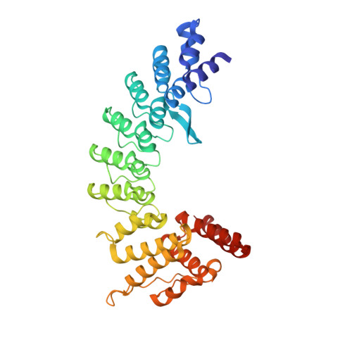
Molecular basis for ubiquitin ligase CRL2 FEM1C -mediated recognition of C-degron.

Yan, X.Wang, X.Li, Y.Zhou, M.Li, Y.Song, L.Mi, W.Min, J.Dong, C.

(2021) Nat Chem Biol 17: 263-271

  • DOI: https://doi.org/10.1038/s41589-020-00703-4
  • Primary Citation of Related Structures:  
    6XKC, 7JYA

  • PubMed Abstract: 

    Proteome integrity depends on the ubiquitin-proteasome system to degrade unwanted or abnormal proteins. In addition to the N-degrons, C-terminal residues of proteins can also serve as degradation signals (C-degrons) that are recognized by specific cullin-RING ubiquitin ligases (CRLs) for proteasomal degradation. FEM1C is a CRL2 substrate receptor that targets the C-terminal arginine degron (Arg/C-degron), but the molecular mechanism of substrate recognition remains largely elusive. Here, we present crystal structures of FEM1C in complex with Arg/C-degron and show that FEM1C utilizes a semi-open binding pocket to capture the C-terminal arginine and that the extreme C-terminal arginine is the major structural determinant in recognition by FEM1C. Together with biochemical and mutagenesis studies, we provide a framework for understanding molecular recognition of the Arg/C-degron by the FEM family of proteins.


  • Organizational Affiliation

    Department of Biochemistry and Molecular Biology, The Province and Ministry Co-sponsored Collaborative Innovation Center for Medical Epigenetics, School of Basic Medical Sciences, Tianjin Medical University, Tianjin, China.


Macromolecules
Find similar proteins by:  (by identity cutoff)  |  3D Structure
Entity ID: 1
MoleculeChains Sequence LengthOrganismDetailsImage
Protein fem-1 homolog C
A, B, C
371Homo sapiensMutation(s): 0 
Gene Names: FEM1CKIAA1785
UniProt & NIH Common Fund Data Resources
Find proteins for Q96JP0 (Homo sapiens)
Explore Q96JP0 
Go to UniProtKB:  Q96JP0
PHAROS:  Q96JP0
GTEx:  ENSG00000145780 
Entity Groups  
Sequence Clusters30% Identity50% Identity70% Identity90% Identity95% Identity100% Identity
UniProt GroupQ96JP0
Sequence Annotations
Expand
  • Reference Sequence

Find similar proteins by:  Sequence   |   3D Structure  

Entity ID: 2
MoleculeChains Sequence LengthOrganismDetailsImage
ASN-ARG-ARG-ARG-ARG-TRP-ARG-GLU-ARG-GLN-ARG
D, E, F
11Human immunodeficiency virus 1Mutation(s): 0 
UniProt
Find proteins for P04618 (Human immunodeficiency virus type 1 group M subtype B (isolate HXB2))
Explore P04618 
Go to UniProtKB:  P04618
Entity Groups  
Sequence Clusters30% Identity50% Identity70% Identity90% Identity95% Identity100% Identity
UniProt GroupP04618
Sequence Annotations
Expand
  • Reference Sequence
Experimental Data & Validation

Experimental Data

  • Method: X-RAY DIFFRACTION
  • Resolution: 2.46 Å
  • R-Value Free: 0.260 
  • R-Value Work: 0.220 
  • R-Value Observed: 0.222 
  • Space Group: P 41 21 2
Unit Cell:
Length ( Å )Angle ( ˚ )
a = 100.133α = 90
b = 100.133β = 90
c = 287.48γ = 90
Software Package:
Software NamePurpose
SCALEPACKdata scaling
PHASERphasing
REFMACrefinement
PDB_EXTRACTdata extraction
HKL-3000data reduction

Structure Validation

View Full Validation Report


Entry History 

Deposition Data

Revision History  (Full details and data files)

  • Version 1.0: 2020-10-14
    Type: Initial release
  • Version 1.1: 2020-12-23
    Changes: Database references
  • Version 1.2: 2021-01-20
    Changes: Database references
  • Version 1.3: 2021-03-10
    Changes: Database references
  • Version 1.4: 2023-10-18
    Changes: Data collection, Database references, Refinement description