7JY9 | pdb_00007jy9

Structure of a 9 base pair RecA-D loop complex


Experimental Data Snapshot

  • Method: ELECTRON MICROSCOPY
  • Resolution: 2.70 Å
  • Aggregation State: PARTICLE 
  • Reconstruction Method: SINGLE PARTICLE 

Starting Model: experimental
View more details

wwPDB Validation 3D Report Full Report

Validation slider image for 7JY9

This is version 1.3 of the entry. See complete history

Literature

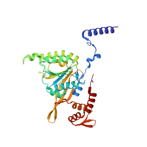
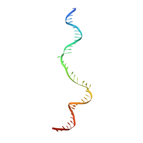
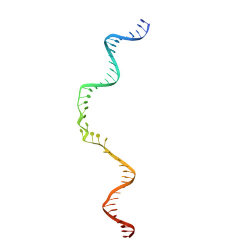
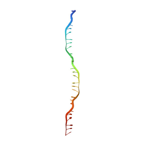
Mechanism of strand exchange from RecA-DNA synaptic and D-loop structures.

Yang, H.Zhou, C.Dhar, A.Pavletich, N.P.

(2020) Nature 586: 801-806

  • DOI: https://doi.org/10.1038/s41586-020-2820-9
  • Primary Citation Related Structures: 
    7JY6, 7JY7, 7JY8, 7JY9

  • PubMed Abstract: 

    The strand-exchange reaction is central to homologous recombination. It is catalysed by the RecA family of ATPases, which form a helical filament with single-stranded DNA (ssDNA) and ATP. This filament binds to a donor double-stranded DNA (dsDNA) to form synaptic filaments, which search for homology and then catalyse the exchange of the complementary strand, forming either a new heteroduplex or-if homology is limited-a D-loop 1,2 . How synaptic filaments form, search for homology and catalyse strand exchange is poorly understood. Here we report the cryo-electron microscopy analysis of synaptic mini-filaments with both non-complementary and partially complementary dsDNA, and structures of RecA-D-loop complexes containing a 10- or a 12-base-pair heteroduplex. The C-terminal domain of RecA binds to dsDNA and directs it to the RecA L2 loop, which inserts into and opens up the duplex. The opening propagates through RecA sequestering the homologous strand at a secondary DNA-binding site, which frees the complementary strand to sample pairing with the ssDNA. At each RecA step, there is a roughly 20% probability that duplex opening will terminate and the as-yet-unopened dsDNA portion will bind to another C-terminal domain. Homology suppresses this process, through the cooperation of heteroduplex pairing with the binding of ssDNA to the secondary site, to extend dsDNA opening. This mechanism locally limits the length of ssDNA sampled for pairing if homology is not encountered, and could allow for the formation of multiple, widely separated synapses on the donor dsDNA, which would increase the likelihood of encountering homology. These findings provide key mechanistic insights into homologous recombination.


  • Organizational Affiliation
    • Structural Biology Program, Memorial Sloan-Kettering Cancer Center, New York, NY, USA.

Macromolecule Content 

  • Total Structure Weight: 364.09 kDa 
  • Atom Count: 24,849 
  • Modeled Residue Count: 3,075 
  • Deposited Residue Count: 3,123 
  • Unique protein chains: 1
  • Unique nucleic acid chains: 3

Macromolecules


Find similar proteins by:|  3D Structure
Entity ID: 1
MoleculeChains  Sequence LengthOrganismDetailsImage
Protein RecA
A, B, C, D, E
A, B, C, D, E, F, G, H, I
334Escherichia coliMutation(s): 0 
Gene Names: recANCTC11341_01072
UniProt
Find proteins for P0A7G6 (Escherichia coli (strain K12))
Explore P0A7G6 
Go to UniProtKB:  P0A7G6
Entity Groups
Sequence Clusters30% Identity50% Identity70% Identity90% Identity95% Identity100% Identity
UniProt GroupP0A7G6
Sequence Annotations
Expand
Reference Sequence
Find similar nucleic acids by:  (by identity cutoff) 
Entity ID: 2
MoleculeChains LengthOrganismImage
DNA (42-MER)J [auth T]45Escherichia coli
Sequence Annotations
Expand
Reference Sequence
Find similar nucleic acids by:  (by identity cutoff) 
Entity ID: 3
MoleculeChains LengthOrganismImage
DNA (42-MER)K [auth U]45Escherichia coli
Sequence Annotations
Expand
Reference Sequence
Find similar nucleic acids by:  (by identity cutoff) 
Entity ID: 4
MoleculeChains LengthOrganismImage
DNA (27-MER)L [auth S]27Escherichia coli
Sequence Annotations
Expand
Reference Sequence

Small Molecules

Ligands 2 Unique
IDChains Name / Formula / InChI Key2D Diagram3D Interactions
AGS

Query on AGS


Download:Ideal Coordinates CCD File
BA [auth H]
DA [auth I]
N [auth A]
P [auth B]
R [auth C]
BA [auth H],
DA [auth I],
N [auth A],
P [auth B],
R [auth C],
T [auth D],
V [auth E],
X [auth F],
Z [auth G]
PHOSPHOTHIOPHOSPHORIC ACID-ADENYLATE ESTER
C10 H16 N5 O12 P3 S
NLTUCYMLOPLUHL-KQYNXXCUSA-N
MG

Query on MG


Download:Ideal Coordinates CCD File
AA [auth H]
CA [auth I]
M [auth A]
O [auth B]
Q [auth C]
AA [auth H],
CA [auth I],
M [auth A],
O [auth B],
Q [auth C],
S [auth D],
U [auth E],
W [auth F],
Y [auth G]
MAGNESIUM ION
Mg
JLVVSXFLKOJNIY-UHFFFAOYSA-N

Experimental Data & Validation

Experimental Data

  • Method: ELECTRON MICROSCOPY
  • Resolution: 2.70 Å
  • Aggregation State: PARTICLE 
  • Reconstruction Method: SINGLE PARTICLE 
EM Software:
TaskSoftware PackageVersion
MODEL REFINEMENTREFMAC

Structure Validation

View Full Validation Report


Entry History 

& Funding Information

Deposition Data


Funding OrganizationLocationGrant Number
Howard Hughes Medical Institute (HHMI)United States--

Revision History  (Full details and data files)

  • Version 1.0: 2020-11-04
    Type: Initial release
  • Version 1.1: 2020-11-11
    Changes: Database references
  • Version 1.2: 2024-03-06
    Changes: Data collection, Database references, Refinement description
  • Version 1.3: 2024-11-20
    Changes: Data collection, Structure summary