7JXF

E. coli TSase complex with a bi-substrate reaction intermediate diastereomer analog


Experimental Data Snapshot

  • Method: X-RAY DIFFRACTION
  • Resolution: 1.50 Å
  • R-Value Free: 0.175 
  • R-Value Work: 0.151 
  • R-Value Observed: 0.152 

wwPDB Validation   3D Report Full Report


Ligand Structure Quality Assessment 


This is version 1.2 of the entry. See complete history


Literature

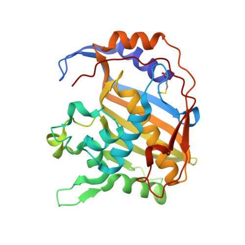
Caught in Action: X-ray Structure of Thymidylate Synthase with Noncovalent Intermediate Analog.

Kholodar, S.A.Finer-Moore, J.S.Swiderek, K.Arafet, K.Moliner, V.Stroud, R.M.Kohen, A.

(2021) Biochemistry 60: 1243-1247

  • DOI: https://doi.org/10.1021/acs.biochem.1c00063
  • Primary Citation of Related Structures:  
    7JX1, 7JXF

  • PubMed Abstract: 

    Methylation of 2-deoxyuridine-5'-monophosphate (dUMP) at the C5 position by the obligate dimeric thymidylate synthase (TSase) in the sole de novo biosynthetic pathway to thymidine 5'-monophosphate (dTMP) proceeds by forming a covalent ternary complex with dUMP and cosubstrate 5,10-methylenetetrahydrofolate. The crystal structure of an analog of this intermediate gives important mechanistic insights but does not explain the half-of-the-sites activity of the enzyme. Recent experiments showed that the C5 proton and the catalytic Cys are eliminated in a concerted manner from the covalent ternary complex to produce a noncovalent bisubstrate intermediate. Here, we report the crystal structure of TSase with a close synthetic analog of this intermediate in which it has partially reacted with the enzyme but in only one protomer, consistent with the half-of-the-sites activity of this enzyme. Quantum mechanics/molecular mechanics simulations confirmed that the analog could undergo catalysis. The crystal structure shows a new water 2.9 Å from the critical C5 of the dUMP moiety, which in conjunction with other residues in the network, may be the elusive general base that abstracts the C5 proton of dUMP during the reaction.


  • Organizational Affiliation

    Department of Chemistry, The University of Iowa, Iowa City, Iowa 52242, United States.


Macromolecules
Find similar proteins by:  (by identity cutoff)  |  3D Structure
Entity ID: 1
MoleculeChains Sequence LengthOrganismDetailsImage
Thymidylate synthase
A, B
264Escherichia coliMutation(s): 0 
Gene Names: thyA
EC: 2.1.1.45
UniProt
Find proteins for P0A884 (Escherichia coli (strain K12))
Explore P0A884 
Go to UniProtKB:  P0A884
Entity Groups  
Sequence Clusters30% Identity50% Identity70% Identity90% Identity95% Identity100% Identity
UniProt GroupP0A884
Sequence Annotations
Expand
  • Reference Sequence
Small Molecules
Ligands 4 Unique
IDChains Name / Formula / InChI Key2D Diagram3D Interactions
VNM (Subject of Investigation/LOI)
Query on VNM

Download Ideal Coordinates CCD File 
G [auth B](2S)-2-({4-[({(6S)-2,4-diamino-5-[(1-{(2R,4S,5R)-4-hydroxy-5-[(phosphonooxy)methyl]tetrahydrofuran-2-yl}-2,4-dioxo-1,2,3,4-tetrahydropyrimidin-5-yl)methyl]-5,6,7,8-tetrahydropyrido[3,2-d]pyrimidin-6-yl}methyl)amino]benzoyl}amino)pentanedioic acid (non-preferred name)
C30 H38 N9 O13 P
OYTZJQBUPHBEFY-GLOJMNGXSA-N
VMV (Subject of Investigation/LOI)
Query on VMV

Download Ideal Coordinates CCD File 
D [auth A]N-[4-({[(6S)-2,4-diamino-5,6,7,8-tetrahydropyrido[3,2-d]pyrimidin-6-yl]methyl}amino)benzene-1-carbonyl]-L-glutamic acid
C20 H25 N7 O5
NJZPEYYRVZHUEO-JSGCOSHPSA-N
5HU (Subject of Investigation/LOI)
Query on 5HU

Download Ideal Coordinates CCD File 
C [auth A]5-HYDROXYMETHYLURIDINE-2'-DEOXY-5'-MONOPHOSPHATE
C10 H15 N2 O9 P
WEBVWKFGRVLCNS-XLPZGREQSA-N
NA (Subject of Investigation/LOI)
Query on NA

Download Ideal Coordinates CCD File 
E [auth A],
F [auth A],
H [auth B]
SODIUM ION
Na
FKNQFGJONOIPTF-UHFFFAOYSA-N
Modified Residues  1 Unique
IDChains TypeFormula2D DiagramParent
CXM
Query on CXM
A, B
L-PEPTIDE LINKINGC6 H11 N O4 SMET
Experimental Data & Validation

Experimental Data

  • Method: X-RAY DIFFRACTION
  • Resolution: 1.50 Å
  • R-Value Free: 0.175 
  • R-Value Work: 0.151 
  • R-Value Observed: 0.152 
  • Space Group: P 63
Unit Cell:
Length ( Å )Angle ( ˚ )
a = 125.442α = 90
b = 125.442β = 90
c = 67.049γ = 120
Software Package:
Software NamePurpose
PHENIXrefinement
XDSdata reduction
XSCALEdata scaling
PDB_EXTRACTdata extraction
PHENIXphasing

Structure Validation

View Full Validation Report


Ligand Structure Quality Assessment 


Entry History & Funding Information

Deposition Data


Funding OrganizationLocationGrant Number
National Institutes of Health/National Institute of General Medical Sciences (NIH/NIGMS)United StatesGM24485

Revision History  (Full details and data files)

  • Version 1.0: 2021-04-21
    Type: Initial release
  • Version 1.1: 2021-05-05
    Changes: Database references
  • Version 1.2: 2023-10-18
    Changes: Data collection, Database references, Refinement description