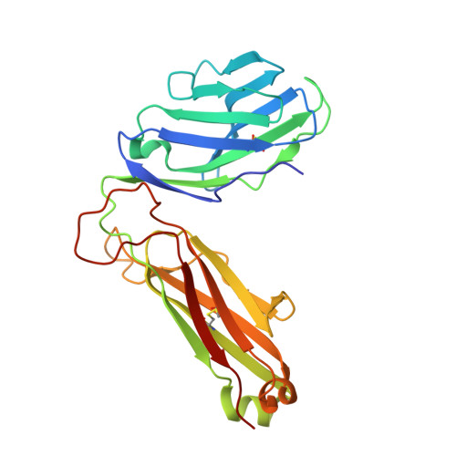
7JWI | pdb_00007jwi

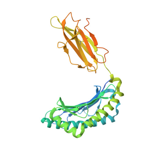
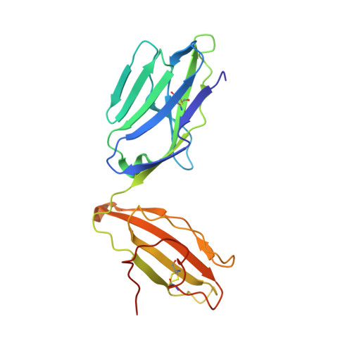
Crystal structure of B17.R2 TCR in complex with H2D-b-NP366


Experimental Data Snapshot

  • Method: X-RAY DIFFRACTION
  • Resolution: 3.02 Å
  • R-Value Free: 
    0.281 (Depositor), 0.296 (DCC) 
  • R-Value Work: 
    0.239 (Depositor), 0.250 (DCC) 

Starting Model: experimental
View more details

wwPDB Validation 3D Report Full Report

Validation slider image for 7JWI

This is version 1.2 of the entry. See complete history. 

Literature

Canonical T cell receptor docking on peptide-MHC is essential for T cell signaling.

Zareie, P., Szeto, C., Farenc, C., Gunasinghe, S.D., Kolawole, E.M., Nguyen, A., Blyth, C., Sng, X.Y.X., Li, J., Jones, C.M., Fulcher, A.J., Jacobs, J.R., Wei, Q., Wojciech, L., Petersen, J., Gascoigne, N.R.J., Evavold, B.D., Gaus, K., Gras, S., Rossjohn, J., La Gruta, N.L.

(2021) Science 372

  • DOI: https://doi.org/10.1126/science.abe9124
  • Primary Citation Related Structures: 
    7JWI, 7JWJ

  • PubMed Abstract: 

    T cell receptor (TCR) recognition of peptide-major histocompatibility complexes (pMHCs) is characterized by a highly conserved docking polarity. Whether this polarity is driven by recognition or signaling constraints remains unclear. Using "reversed-docking" TCRβ-variable (TRBV) 17 + TCRs from the naïve mouse CD8 + T cell repertoire that recognizes the H-2D b -NP 366 epitope, we demonstrate that their inability to support T cell activation and in vivo recruitment is a direct consequence of reversed docking polarity and not TCR-pMHCI binding or clustering characteristics. Canonical TCR-pMHCI docking optimally localizes CD8/Lck to the CD3 complex, which is prevented by reversed TCR-pMHCI polarity. The requirement for canonical docking was circumvented by dissociating Lck from CD8. Thus, the consensus TCR-pMHC docking topology is mandated by T cell signaling constraints.


  • Organizational Affiliation: 
    • Infection and Immunity Program and Department of Biochemistry and Molecular Biology, Biomedicine Discovery Institute, Monash University, Clayton, Victoria, Australia.

Macromolecule Content 

  • Total Structure Weight: 104.84 kDa 
  • Atom Count: 6,864 
  • Modeled Residue Count: 828 
  • Deposited Residue Count: 921 
  • Unique protein chains: 5

Macromolecules

Find similar proteins by:|  3D Structure
Entity ID: 1
MoleculeChains  Sequence LengthOrganismDetailsImage
H-2 class I histocompatibility antigen, D-B alpha chain362Mus musculusMutation(s): 0 
Gene Names: H2-D1
UniProt
Find proteins for P01899 (Mus musculus)
Explore P01899 
Go to UniProtKB:  P01899
Entity Groups
Sequence Clusters30% Identity50% Identity70% Identity90% Identity95% Identity100% Identity
UniProt GroupP01899
Sequence Annotations
Expand
Reference Sequence
Find similar proteins by:|  3D Structure
Entity ID: 2
MoleculeChains  Sequence LengthOrganismDetailsImage
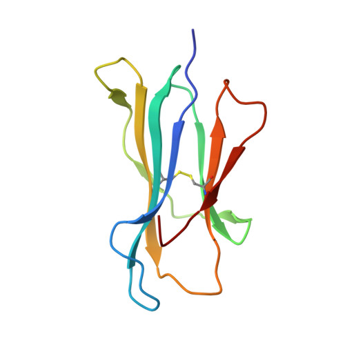
Beta-2-microglobulin100Homo sapiensMutation(s): 0 
Gene Names: B2M, CDABP0092, HDCMA22P
UniProt & NIH Common Fund Data Resources
Find proteins for P61769 (Homo sapiens)
Explore P61769 
Go to UniProtKB:  P61769
PHAROS:  P61769
GTEx:  ENSG00000166710 
Entity Groups
Sequence Clusters30% Identity50% Identity70% Identity90% Identity95% Identity100% Identity
UniProt GroupP61769
Sequence Annotations
Expand
Reference Sequence
Find similar proteins by:  Sequence   |   3D Structure  
Entity ID: 3
MoleculeChains  Sequence LengthOrganismDetailsImage
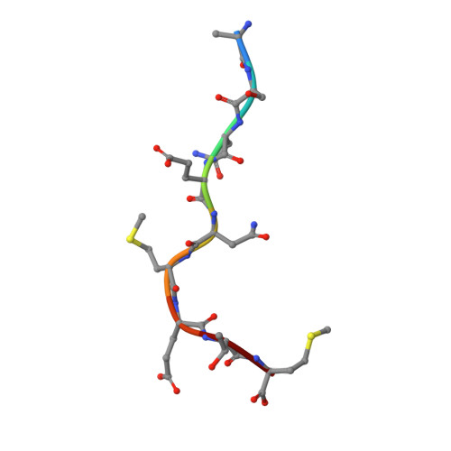
Nucleoprotein9Influenza A virusMutation(s): 0 
UniProt
Find proteins for Q9Q0U8 (Influenza A virus (strain A/Goose/Guangdong/1/1996 H5N1 genotype Gs/Gd))
Explore Q9Q0U8 
Go to UniProtKB:  Q9Q0U8
Entity Groups
UniProt GroupQ9Q0U8
Sequence Annotations
Expand
Reference Sequence
Find similar proteins by:|  3D Structure
Entity ID: 4
MoleculeChains  Sequence LengthOrganismDetailsImage
B17.R2 TCR alpha chain207Mus musculusMutation(s): 0 
Entity Groups
Sequence Clusters30% Identity50% Identity70% Identity90% Identity95% Identity100% Identity
Sequence Annotations
Expand
Reference Sequence
Find similar proteins by:|  3D Structure
Entity ID: 5
MoleculeChains  Sequence LengthOrganismDetailsImage
B17.R2 TCR beta chain243Mus musculusMutation(s): 0 
Entity Groups
Sequence Clusters30% Identity50% Identity70% Identity90% Identity95% Identity100% Identity
Sequence Annotations
Expand
Reference Sequence

Small Molecules

Ligands 1 Unique
IDChains Name / Formula / InChI Key2D Diagram3D Interactions
EDO

Query on EDO


Download:Ideal Coordinates CCD File
F [auth E]1,2-ETHANEDIOL
C2 H6 O2
LYCAIKOWRPUZTN-UHFFFAOYSA-N

Experimental Data & Validation

Experimental Data

  • Method: X-RAY DIFFRACTION
  • Resolution: 3.02 Å
  • R-Value Free:  0.281 (Depositor), 0.296 (DCC) 
  • R-Value Work:  0.239 (Depositor), 0.250 (DCC) 
Space Group: I 2 2 2
Unit Cell:
Length ( Å )Angle ( ˚ )
a = 91.668α = 90
b = 140.997β = 90
c = 217.649γ = 90
Software Package:
Software NamePurpose
BUSTERrefinement
PDB_EXTRACTdata extraction
XFITdata reduction
Aimlessdata scaling
PHASERphasing

Structure Validation

View Full Validation Report


Entry History 

Deposition Data

Revision History  (Full details and data files)

  • Version 1.0: 2021-07-07
    Type: Initial release
  • Version 1.1: 2023-10-18
    Changes: Data collection, Database references, Refinement description
  • Version 1.2: 2024-10-09
    Changes: Structure summary