7JVL

Structure of the M101A variant of the SidA ornithine hydroxylase complexed with NADP and the FAD in the "out" conformation


Experimental Data Snapshot

  • Method: X-RAY DIFFRACTION
  • Resolution: 2.10 Å
  • R-Value Free: 0.241 
  • R-Value Work: 0.204 
  • R-Value Observed: 0.205 

wwPDB Validation   3D Report Full Report


Ligand Structure Quality Assessment 


This is version 1.2 of the entry. See complete history


Literature

Structural Determinants of Flavin Dynamics in a Class B Monooxygenase.

Campbell, A.C.Robinson, R.Mena-Aguilar, D.Sobrado, P.Tanner, J.J.

(2020) Biochemistry 59: 4609-4616

  • DOI: https://doi.org/10.1021/acs.biochem.0c00783
  • Primary Citation of Related Structures:  
    7JVK, 7JVL

  • PubMed Abstract: 

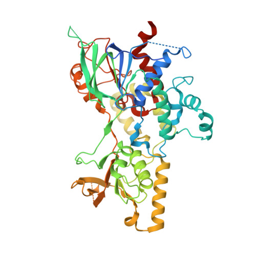
    The ornithine hydroxylase known as SidA is a class B flavin monooxygenase that catalyzes the first step in the biosynthesis of hydroxamate-containing siderophores in Aspergillus fumigatus . Crystallographic studies of SidA revealed that the FAD undergoes dramatic conformational changes between out and in states during the catalytic cycle. We sought insight into the origins and purpose of flavin motion in class B monooxygenases by probing the function of Met101, a residue that contacts the pyrimidine ring of the in FAD. Steady-state kinetic measurements showed that the mutant variant M101A has a 25-fold lower turnover number. Pre-steady-state kinetic measurements, pH profiles, and solvent kinetic isotope effect measurements were used to isolate the microscopic step that is responsible for the reduced steady-state activity. The data are consistent with a bottleneck in the final step of the mechanism, which involves flavin dehydration and the release of hydroxy-l-ornithine and NADP + . Crystal structures were determined for M101A in the resting state and complexed with NADP + . The resting enzyme structure is similar to that of wild-type SidA, consistent with M101A exhibiting normal kinetics for flavin reduction by NADPH and wild-type affinity for NADPH. In contrast, the structure of the M101A-NADP + complex unexpectedly shows the FAD adopting the out conformation and may represent a stalled conformation that is responsible for the slow kinetics. Altogether, our data support a previous proposal that one purpose of the FAD conformational change from in to out in class B flavin monooxygenases is to eject spent NADP + in preparation for a new catalytic cycle.


  • Organizational Affiliation

    Department of Biochemistry, University of Missouri, Columbia, Missouri 65211, United States.


Macromolecules
Find similar proteins by:  (by identity cutoff)  |  3D Structure
Entity ID: 1
MoleculeChains Sequence LengthOrganismDetailsImage
L-ornithine N(5)-monooxygenase
A, B, C, D
501Aspergillus fumigatus Af293Mutation(s): 1 
Gene Names: sidAAfu2g07680
EC: 1.14.13.196
UniProt
Find proteins for E9QYP0 (Aspergillus fumigatus (strain ATCC MYA-4609 / CBS 101355 / FGSC A1100 / Af293))
Explore E9QYP0 
Go to UniProtKB:  E9QYP0
Entity Groups  
Sequence Clusters30% Identity50% Identity70% Identity90% Identity95% Identity100% Identity
UniProt GroupE9QYP0
Sequence Annotations
Expand
  • Reference Sequence
Small Molecules
Ligands 4 Unique
IDChains Name / Formula / InChI Key2D Diagram3D Interactions
FAD (Subject of Investigation/LOI)
Query on FAD

Download Ideal Coordinates CCD File 
E [auth A],
I [auth B],
N [auth C],
R [auth D]
FLAVIN-ADENINE DINUCLEOTIDE
C27 H33 N9 O15 P2
VWWQXMAJTJZDQX-UYBVJOGSSA-N
NAP (Subject of Investigation/LOI)
Query on NAP

Download Ideal Coordinates CCD File 
F [auth A],
J [auth B],
O [auth C],
S [auth D]
NADP NICOTINAMIDE-ADENINE-DINUCLEOTIDE PHOSPHATE
C21 H28 N7 O17 P3
XJLXINKUBYWONI-NNYOXOHSSA-N
ACT
Query on ACT

Download Ideal Coordinates CCD File 
G [auth A]
H [auth A]
K [auth B]
L [auth B]
P [auth C]
G [auth A],
H [auth A],
K [auth B],
L [auth B],
P [auth C],
Q [auth C],
T [auth D],
U [auth D]
ACETATE ION
C2 H3 O2
QTBSBXVTEAMEQO-UHFFFAOYSA-M
CA
Query on CA

Download Ideal Coordinates CCD File 
M [auth B],
V [auth D]
CALCIUM ION
Ca
BHPQYMZQTOCNFJ-UHFFFAOYSA-N
Experimental Data & Validation

Experimental Data

  • Method: X-RAY DIFFRACTION
  • Resolution: 2.10 Å
  • R-Value Free: 0.241 
  • R-Value Work: 0.204 
  • R-Value Observed: 0.205 
  • Space Group: P 1 21 1
Unit Cell:
Length ( Å )Angle ( ˚ )
a = 80.229α = 90
b = 153.811β = 109.28
c = 89.87γ = 90
Software Package:
Software NamePurpose
Aimlessdata scaling
PHENIXrefinement
PDB_EXTRACTdata extraction
XDSdata reduction
PHENIXphasing

Structure Validation

View Full Validation Report


Ligand Structure Quality Assessment 


Entry History & Funding Information

Deposition Data


Funding OrganizationLocationGrant Number
National Science Foundation (NSF, United States)United StatesCHE-2003658
National Science Foundation (NSF, United States)United StatesCHE-2003986

Revision History  (Full details and data files)

  • Version 1.0: 2020-12-02
    Type: Initial release
  • Version 1.1: 2020-12-16
    Changes: Database references
  • Version 1.2: 2023-10-18
    Changes: Data collection, Database references, Refinement description