Structures of an engineered phospholipase D with specificity for secondary alcohol transphosphatidylation: insights into plasticity of substrate binding and activation.
Samantha, A., Damnjanovic, J., Iwasaki, Y., Nakano, H., Vrielink, A.(2021) Biochem J 478: 1749-1767
- PubMed: 33843991 Search on PubMedSearch on PubMed Central
- DOI: https://doi.org/10.1042/BCJ20210117
- Primary Citation Related Structures:
7JRB, 7JRC, 7JRU, 7JRV, 7JRW, 7JS5, 7JS7 - PubMed Abstract:
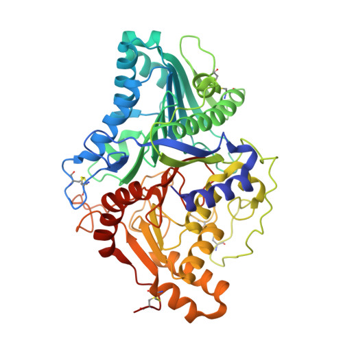
Phospholipase D (PLD) is an enzyme useful for the enzymatic modification of phospholipids. In the presence of primary alcohols, the enzyme catalyses transphosphatidylation of the head group of phospholipid substrates to synthesise a modified phospholipid product. However, the enzyme is specific for primary alcohols and thus the limitation of the molecular size of the acceptor compounds has restricted the type of phospholipid species that can be synthesised. An engineered variant of PLD from Streptomyces antibioticus termed TNYR SaPLD was developed capable of synthesising 1-phosphatidylinositol with positional specificity of up to 98%. To gain a better understanding of the substrate binding features of the TNYR SaPLD, crystal structures have been determined for the free enzyme and its complexes with phosphate, phosphatidic acid and 1-inositol phosphate. Comparisons of these structures with the wild-type SaPLD show a larger binding site able to accommodate a bulkier secondary alcohol substrate as well as changes to the position of a flexible surface loop proposed to be involved in substrate recognition. The complex of the active TNYR SaPLD with 1-inositol phosphate reveals a covalent intermediate adduct with the ligand bound to H442 rather than to H168, the proposed nucleophile in the wild-type enzyme. This structural feature suggests that the enzyme exhibits plasticity of the catalytic mechanism different from what has been reported to date for PLDs. These structural studies provide insights into the underlying mechanism that governs the recognition of myo-inositol by TNYR SaPLD, and an important foundation for further studies of the catalytic mechanism.
- School of Molecular Sciences, University of Western Australia, 35 Stirling Highway, Crawley, WA 6009, Australia.
Organizational Affiliation: