7JQG

Crystal structure of human PPARgamma ligand binding domain Y473E mutant in complex with GW1929


Experimental Data Snapshot

  • Method: X-RAY DIFFRACTION
  • Resolution: 2.15 Å
  • R-Value Free: 0.270 
  • R-Value Work: 0.225 
  • R-Value Observed: 0.228 

wwPDB Validation   3D Report Full Report


Ligand Structure Quality Assessment 


This is version 1.2 of the entry. See complete history


Literature

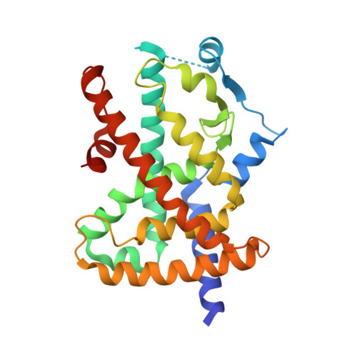
Structural mechanism underlying ligand binding and activation of PPAR gamma.

Shang, J.Kojetin, D.J.

(2021) Structure 29: 940-950.e4

  • DOI: https://doi.org/10.1016/j.str.2021.02.006
  • Primary Citation of Related Structures:  
    6VZL, 6VZM, 6VZN, 6VZO, 7JQG

  • PubMed Abstract: 

    Ligands bind to an occluded orthosteric ligand-binding pocket within the nuclear receptor ligand-binding domain. Molecular simulations have revealed theoretical ligand entry/exit pathways to the orthosteric pocket; however, it remains unclear whether ligand binding proceeds through induced fit or conformational selection mechanisms. Here, using nuclear magnetic resonance spectroscopy, isothermal titration calorimetry, and surface plasmon resonance analysis, we provide evidence that structurally distinct agonists bind peroxisome proliferator-activated receptor γ (PPARγ) via a two-step induced fit mechanism involving an initial fast kinetic step followed by a slow conformational change. The agonist encounter complex binding pose is suggested in crystal structures where ligands bind to a surface pore suggested as a ligand entry site in molecular simulations. Our findings suggest an activation mechanism for PPARγ whereby agonist binding occurs through an initial encounter complex followed by a transition of the ligand into the final binding pose within the orthosteric pocket, inducing a transcriptionally active conformation.


  • Organizational Affiliation

    Department of Integrative Structural and Computational Biology, The Scripps Research Institute, Jupiter, FL 33458, USA.


Macromolecules
Find similar proteins by:  (by identity cutoff)  |  3D Structure
Entity ID: 1
MoleculeChains Sequence LengthOrganismDetailsImage
Peroxisome proliferator-activated receptor gamma
A, B
276Homo sapiensMutation(s): 1 
Gene Names: PPARGNR1C3
UniProt & NIH Common Fund Data Resources
Find proteins for P37231 (Homo sapiens)
Explore P37231 
Go to UniProtKB:  P37231
PHAROS:  P37231
GTEx:  ENSG00000132170 
Entity Groups  
Sequence Clusters30% Identity50% Identity70% Identity90% Identity95% Identity100% Identity
UniProt GroupP37231
Sequence Annotations
Expand
  • Reference Sequence
Small Molecules
Ligands 1 Unique
IDChains Name / Formula / InChI Key2D Diagram3D Interactions
EDK (Subject of Investigation/LOI)
Query on EDK

Download Ideal Coordinates CCD File 
C [auth A],
D [auth B]
(2~{S})-3-[4-[2-[methyl(pyridin-2-yl)amino]ethoxy]phenyl]-2-[[2-(phenylcarbonyl)phenyl]amino]propanoic acid
C30 H29 N3 O4
QTQMRBZOBKYXCG-MHZLTWQESA-N
Experimental Data & Validation

Experimental Data

  • Method: X-RAY DIFFRACTION
  • Resolution: 2.15 Å
  • R-Value Free: 0.270 
  • R-Value Work: 0.225 
  • R-Value Observed: 0.228 
  • Space Group: C 1 2 1
Unit Cell:
Length ( Å )Angle ( ˚ )
a = 92.699α = 90
b = 61.625β = 102.44
c = 118.66γ = 90
Software Package:
Software NamePurpose
SCALAdata scaling
PHENIXrefinement
PDB_EXTRACTdata extraction
DIALSdata reduction
PHASERphasing

Structure Validation

View Full Validation Report


Ligand Structure Quality Assessment 


Entry History & Funding Information

Deposition Data


Funding OrganizationLocationGrant Number
National Institutes of Health/National Institute of Diabetes and Digestive and Kidney Disease (NIH/NIDDK)United StatesR01DK101871

Revision History  (Full details and data files)

  • Version 1.0: 2021-03-03
    Type: Initial release
  • Version 1.1: 2021-09-29
    Changes: Database references
  • Version 1.2: 2023-10-18
    Changes: Data collection, Refinement description