7JPV | pdb_00007jpv

Rabbit Cav1.1 in the presence of 1 micromolar (S)-(-)-Bay K8644 in nanodiscs at 3.4 Angstrom resolution


Experimental Data Snapshot

  • Method: ELECTRON MICROSCOPY
  • Resolution: 3.40 Å
  • Aggregation State: PARTICLE 
  • Reconstruction Method: SINGLE PARTICLE 

wwPDB Validation 3D Report Full Report

Validation slider image for 7JPV

This is version 1.4 of the entry. See complete history

Literature

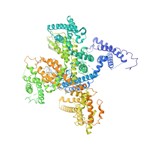
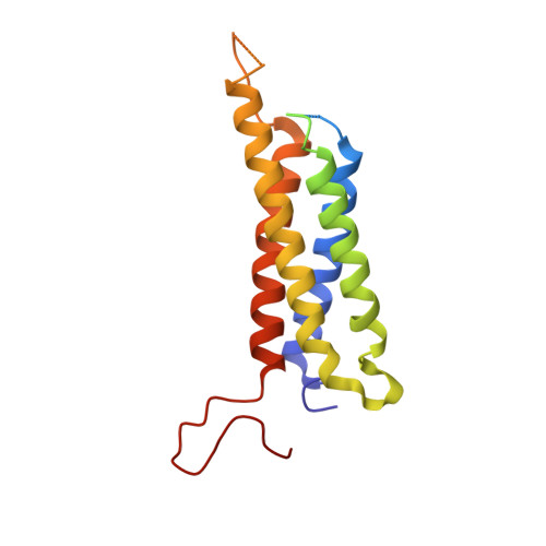
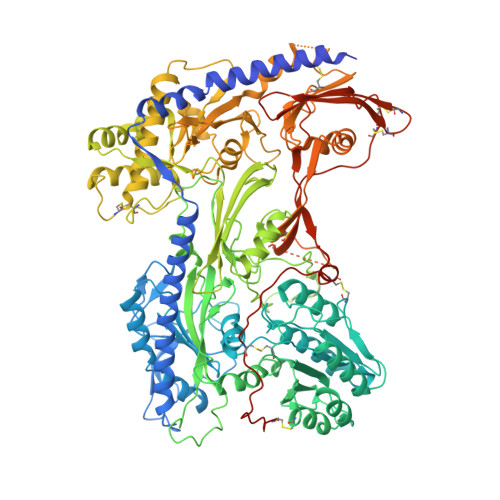
Structural Basis of the Modulation of the Voltage-Gated Calcium Ion Channel Ca v 1.1 by Dihydropyridine Compounds*.

Gao, S.Yan, N.

(2021) Angew Chem Int Ed Engl 60: 3131-3137

  • DOI: https://doi.org/10.1002/anie.202011793
  • Primary Citation Related Structures: 
    7JPK, 7JPL, 7JPV, 7JPW, 7JPX

  • PubMed Abstract: 

    1,4-Dihydropyridines (DHP), the most commonly used antihypertensives, function by inhibiting the L-type voltage-gated Ca 2+ (Ca v ) channels. DHP compounds exhibit chirality-specific antagonistic or agonistic effects. The structure of rabbit Ca v 1.1 bound to an achiral drug nifedipine reveals the general binding mode for DHP drugs, but the molecular basis for chiral specificity remained elusive. Herein, we report five cryo-EM structures of nanodisc-embedded Ca v 1.1 in the presence of the bestselling drug amlodipine, a DHP antagonist (R)-(+)-Bay K8644, and a titration of its agonistic enantiomer (S)-(-)-Bay K8644 at resolutions of 2.9-3.4 Å. The amlodipine-bound structure reveals the molecular basis for the high efficacy of the drug. All structures with the addition of the Bay K8644 enantiomers exhibit similar inactivated conformations, suggesting that (S)-(-)-Bay K8644, when acting as an agonist, is insufficient to lock the activated state of the channel for a prolonged duration.


  • Organizational Affiliation
    • Department of Molecular Biology, Princeton University, Princeton, NJ, 08544, USA.

Macromolecule Content 

  • Total Structure Weight: 369.97 kDa 
  • Atom Count: 18,288 
  • Modeled Residue Count: 2,227 
  • Deposited Residue Count: 3,200 
  • Unique protein chains: 3

Macromolecules

Find similar proteins by:|  3D Structure
Entity ID: 1
MoleculeChains  Sequence LengthOrganismDetailsImage
Voltage-dependent L-type calcium channel subunit alpha-1S1,873Oryctolagus cuniculusMutation(s): 0 
Membrane Entity: Yes 
UniProt
Find proteins for P07293 (Oryctolagus cuniculus)
Explore P07293 
Go to UniProtKB:  P07293
Entity Groups
Sequence Clusters30% Identity50% Identity70% Identity90% Identity95% Identity100% Identity
UniProt GroupP07293
Sequence Annotations
Expand
Reference Sequence
Find similar proteins by:|  3D Structure
Entity ID: 2
MoleculeChains  Sequence LengthOrganismDetailsImage
Voltage-dependent calcium channel gamma-1 subunitB [auth E]222Oryctolagus cuniculusMutation(s): 0 
Membrane Entity: Yes 
UniProt
Find proteins for P19518 (Oryctolagus cuniculus)
Explore P19518 
Go to UniProtKB:  P19518
Entity Groups
Sequence Clusters30% Identity50% Identity70% Identity90% Identity95% Identity100% Identity
UniProt GroupP19518
Sequence Annotations
Expand
Reference Sequence
Find similar proteins by:|  3D Structure
Entity ID: 3
MoleculeChains  Sequence LengthOrganismDetailsImage
Voltage-dependent calcium channel subunit alpha-2/delta-1C [auth F]1,105Oryctolagus cuniculusMutation(s): 0 
Membrane Entity: Yes 
UniProt
Find proteins for P13806 (Oryctolagus cuniculus)
Explore P13806 
Go to UniProtKB:  P13806
Entity Groups
Sequence Clusters30% Identity50% Identity70% Identity90% Identity95% Identity100% Identity
UniProt GroupP13806
Sequence Annotations
Expand
Reference Sequence

Small Molecules

Ligands 3 Unique
IDChains Name / Formula / InChI Key2D Diagram3D Interactions
PC1

Query on PC1


Download:Ideal Coordinates CCD File
N [auth E]1,2-DIACYL-SN-GLYCERO-3-PHOSPHOCHOLINE
C44 H88 N O8 P
NRJAVPSFFCBXDT-HUESYALOSA-N
3PE

Query on 3PE


Download:Ideal Coordinates CCD File
E [auth A]
F [auth A]
G [auth A]
H [auth A]
I [auth A]
E [auth A],
F [auth A],
G [auth A],
H [auth A],
I [auth A],
J [auth A],
K [auth A],
L [auth A],
M [auth A]
1,2-Distearoyl-sn-glycerophosphoethanolamine
C41 H82 N O8 P
LVNGJLRDBYCPGB-LDLOPFEMSA-N
CA

Query on CA


Download:Ideal Coordinates CCD File
D [auth A]CALCIUM ION
Ca
BHPQYMZQTOCNFJ-UHFFFAOYSA-N

Experimental Data & Validation

Experimental Data

  • Method: ELECTRON MICROSCOPY
  • Resolution: 3.40 Å
  • Aggregation State: PARTICLE 
  • Reconstruction Method: SINGLE PARTICLE 
EM Software:
TaskSoftware PackageVersion
MODEL REFINEMENTPHENIX

Structure Validation

View Full Validation Report


Entry History 

& Funding Information

Deposition Data


Funding OrganizationLocationGrant Number
National Science Foundation (NSF, United States)United StatesDMR-1420541

Revision History  (Full details and data files)

  • Version 1.0: 2020-11-18
    Type: Initial release
  • Version 1.1: 2020-12-23
    Changes: Database references
  • Version 1.2: 2021-03-10
    Changes: Database references
  • Version 1.3: 2024-10-23
    Changes: Data collection, Database references, Derived calculations, Structure summary
  • Version 1.4: 2025-05-28
    Changes: Data collection