7JOV | pdb_00007jov

CRYSTAL STRUCTURE OF RHO-ASSOCIATED PROTEIN KINASE 2 (ROCK2) IN COMPLEX WITH A PHENYLPYRAZOLE AMIDE INHIBITOR


Experimental Data Snapshot

  • Method: X-RAY DIFFRACTION
  • Resolution: 2.59 Å
  • R-Value Free: 
    0.217 (Depositor), 0.234 (DCC) 
  • R-Value Work: 
    0.184 (Depositor), 0.199 (DCC) 
  • R-Value Observed: 
    0.185 (Depositor) 

wwPDB Validation 3D Report Full Report

Validation slider image for 7JOV

Ligand Structure Quality Assessment 


This is version 1.1 of the entry. See complete history

Literature

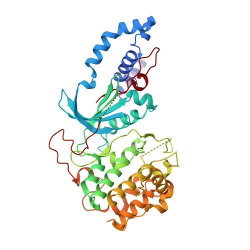
Discovery of a phenylpyrazole amide ROCK inhibitor as a tool molecule for in vivo studies.

Hu, Z.Wang, C.Glunz, P.W.Li, J.Cheadle, N.L.Chen, A.Y.Chen, X.Q.Myers, J.E.Guarino, V.R.Rose, A.Sack, J.S.Sitkoff, D.Taylor, D.S.Xu, S.Yan, C.Zhang, H.Zhang, L.Hennan, J.Adam, L.P.Wexler, R.R.Quan, M.L.

(2020) Bioorg Med Chem Lett 30: 127495-127495

  • DOI: https://doi.org/10.1016/j.bmcl.2020.127495
  • Primary Citation Related Structures: 
    7JOU, 7JOV

  • PubMed Abstract: 

    Structure-activity relationship optimization on a series of phenylpyrazole amides led to the identification of a dual ROCK1 and ROCK2 inhibitor (25) which demonstrated good potency, kinome selectivity and favorable pharmacokinetic profiles. Compound 25 was selected as a tool molecule for in vivo studies including evaluating hemodynamic effects in telemeterized mice, from which moderate decreases in blood pressure were observed.


  • Organizational Affiliation
    • Research & Early Development, Bristol Myers Squibb, P.O. Box 5400, Princeton, NJ 08543-5400, USA. Electronic address: zilun.hu@bms.com.

Macromolecule Content 

  • Total Structure Weight: 366.4 kDa 
  • Atom Count: 23,825 
  • Modeled Residue Count: 3,049 
  • Deposited Residue Count: 3,160 
  • Unique protein chains: 1

Macromolecules

Find similar proteins by:|  3D Structure
Entity ID: 1
MoleculeChains  Sequence LengthOrganismDetailsImage
Rho-associated protein kinase 2
A, B, C, D, E
A, B, C, D, E, F, G, H
395Homo sapiensMutation(s): 0 
Gene Names: ROCK2KIAA0619
EC: 2.7.11.1 (PDB Primary Data), 2.7.11.39 (UniProt)
UniProt & NIH Common Fund Data Resources
Find proteins for O75116 (Homo sapiens)
Explore O75116 
Go to UniProtKB:  O75116
PHAROS:  O75116
GTEx:  ENSG00000134318 
Entity Groups
Sequence Clusters30% Identity50% Identity70% Identity90% Identity95% Identity100% Identity
UniProt GroupO75116
Sequence Annotations
Expand
Reference Sequence

Small Molecules

Ligands 3 Unique
IDChains Name / Formula / InChI Key2D Diagram3D Interactions
VFS
(Subject of Investigation/LOI)

Query on VFS


Download:Ideal Coordinates CCD File
J [auth A]
K [auth B]
M [auth C]
O [auth D]
P [auth E]
J [auth A],
K [auth B],
M [auth C],
O [auth D],
P [auth E],
R [auth F],
T [auth G],
U [auth H]
N-[(1S)-2-hydroxy-1-phenylethyl]-3-methoxy-4-(1H-pyrazol-4-yl)benzamide
C19 H19 N3 O3
CXWOMMUYWIPPCD-QGZVFWFLSA-N
MES

Query on MES


Download:Ideal Coordinates CCD File
I [auth A],
Q [auth F]
2-(N-MORPHOLINO)-ETHANESULFONIC ACID
C6 H13 N O4 S
SXGZJKUKBWWHRA-UHFFFAOYSA-N
CL

Query on CL


Download:Ideal Coordinates CCD File
L [auth B],
N [auth C],
S [auth F]
CHLORIDE ION
Cl
VEXZGXHMUGYJMC-UHFFFAOYSA-M

Experimental Data & Validation

Experimental Data

  • Method: X-RAY DIFFRACTION
  • Resolution: 2.59 Å
  • R-Value Free:  0.217 (Depositor), 0.234 (DCC) 
  • R-Value Work:  0.184 (Depositor), 0.199 (DCC) 
  • R-Value Observed: 0.185 (Depositor) 
Space Group: P 1
Unit Cell:
Length ( Å )Angle ( ˚ )
a = 95.157α = 83.5
b = 101.589β = 73.77
c = 102.973γ = 77.69
Software Package:
Software NamePurpose
XDSdata reduction
Aimlessdata scaling
BUSTERrefinement

Structure Validation

View Full Validation Report


Ligand Structure Quality Assessment 


Entry History 

Deposition Data

Revision History  (Full details and data files)

  • Version 1.0: 2020-09-02
    Type: Initial release
  • Version 1.1: 2024-03-06
    Changes: Data collection, Database references