An autoinhibitory role for the GRF zinc finger domain of DNA glycosylase NEIL3.
Rodriguez, A.A., Wojtaszek, J.L., Greer, B.H., Haldar, T., Gates, K.S., Williams, R.S., Eichman, B.F.(2020) J Biological Chem 295: 15566-15575
- PubMed: 32878989 Search on PubMedSearch on PubMed Central
- DOI: https://doi.org/10.1074/jbc.RA120.015541
- Primary Citation Related Structures:
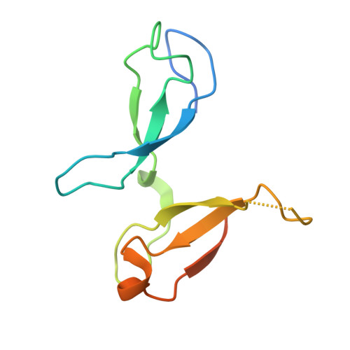
7JL5 - PubMed Abstract:
The NEIL3 DNA glycosylase maintains genome integrity during replication by excising oxidized bases from single-stranded DNA (ssDNA) and unhooking interstrand cross-links (ICLs) at fork structures. In addition to its N-terminal catalytic glycosylase domain, NEIL3 contains two tandem C-terminal GRF-type zinc fingers that are absent in the other NEIL paralogs. ssDNA binding by the GRF-ZF motifs helps recruit NEIL3 to replication forks converged at an ICL, but the nature of DNA binding and the effect of the GRF-ZF domain on catalysis of base excision and ICL unhooking is unknown. Here, we show that the tandem GRF-ZFs of NEIL3 provide affinity and specificity for DNA that is greater than each individual motif alone. The crystal structure of the GRF domain shows that the tandem ZF motifs adopt a flexible head-to-tail configuration well-suited for binding to multiple ssDNA conformations. Functionally, we establish that the NEIL3 GRF domain inhibits glycosylase activity against monoadducts and ICLs. This autoinhibitory activity contrasts GRF-ZF domains of other DNA-processing enzymes, which typically use ssDNA binding to enhance catalytic activity, and suggests that the C-terminal region of NEIL3 is involved in both DNA damage recruitment and enzymatic regulation.
- Department of Biological Sciences and Center for Structural Biology, Vanderbilt University, Nashville, Tennessee, USA.
Organizational Affiliation: