


Crystal structure of the sterol 14alpha-demethylase-ferredoxin (CYP51-fx) heme domain and architectural comparison to the whole fusion protein
Zhao, B., Lamb, D.C.To be published.
Experimental Data Snapshot
Starting Model: experimental
View more details
Entity ID: 1 | |||||
|---|---|---|---|---|---|
| Molecule | Chains | Sequence Length | Organism | Details | Image |
| Cytochrome P450 51 | 450 | Methylococcus capsulatus | Mutation(s): 0 Gene Names: cyp51 EC: 1.14.13.70 | 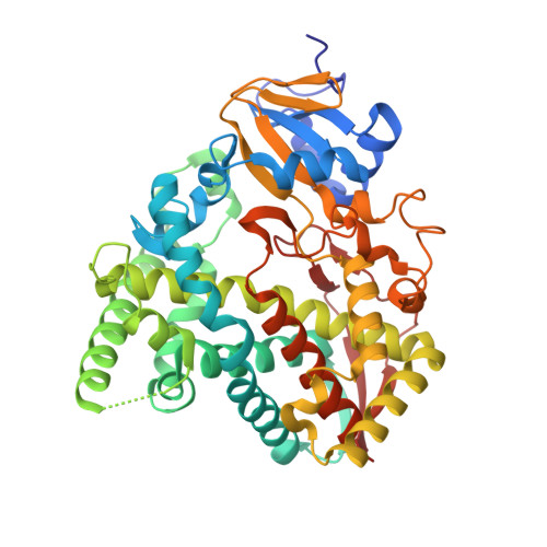 | |
UniProt | |||||
Entity Groups | |||||
| Sequence Clusters | 30% Identity50% Identity70% Identity90% Identity95% Identity100% Identity | ||||
| UniProt Group | Q603T8 | ||||
Sequence AnnotationsExpand | |||||
Reference Sequence | |||||
| Ligands 3 Unique | |||||
|---|---|---|---|---|---|
| ID | Chains | Name / Formula / InChI Key | 2D Diagram | 3D Interactions | |
| HEM (Subject of Investigation/LOI) Download:Ideal Coordinates CCD File | D [auth A], I [auth B], L [auth C] | PROTOPORPHYRIN IX CONTAINING FE C34 H32 Fe N4 O4 KABFMIBPWCXCRK-RGGAHWMASA-L |  | ||
| 16A Download:Ideal Coordinates CCD File | E [auth A], J [auth B], M [auth C] | CETYL-TRIMETHYL-AMMONIUM C19 H42 N RLGQACBPNDBWTB-UHFFFAOYSA-N |  | ||
| IMD Download:Ideal Coordinates CCD File | F [auth A] G [auth A] H [auth A] K [auth B] N [auth C] | IMIDAZOLE C3 H5 N2 RAXXELZNTBOGNW-UHFFFAOYSA-O |  | ||
| Length ( Å ) | Angle ( ˚ ) |
|---|---|
| a = 157.682 | α = 90 |
| b = 164.755 | β = 90 |
| c = 187.683 | γ = 90 |
| Software Name | Purpose |
|---|---|
| PHENIX | refinement |
| PDB_EXTRACT | data extraction |
| HKL-2000 | data reduction |
| HKL-2000 | data scaling |
| PHASER | phasing |
| Funding Organization | Location | Grant Number |
|---|---|---|
| Wellcome Trust | United Kingdom | -- |


