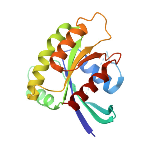
Regulation of GTPase function by autophosphorylation.
Johnson, C.W., Seo, H.S., Terrell, E.M., Yang, M.H., KleinJan, F., Gebregiworgis, T., Gasmi-Seabrook, G.M.C., Geffken, E.A., Lakhani, J., Song, K., Bashyal, P., Popow, O., Paulo, J.A., Liu, A., Mattos, C., Marshall, C.B., Ikura, M., Morrison, D.K., Dhe-Paganon, S., Haigis, K.M.(2022) Mol Cell 82: 950
- PubMed: 35202574 Search on PubMedSearch on PubMed Central
- DOI: https://doi.org/10.1016/j.molcel.2022.02.011
- Primary Citation Related Structures:
7JIF, 7JIG, 7JIH, 7JII, 7KMR - PubMed Abstract:
A unifying feature of the RAS superfamily is a conserved GTPase cycle by which these proteins transition between active and inactive states. We demonstrate that autophosphorylation of some GTPases is an intrinsic regulatory mechanism that reduces nucleotide hydrolysis and enhances nucleotide exchange, altering the on/off switch that forms the basis for their signaling functions. Using X-ray crystallography, nuclear magnetic resonance spectroscopy, binding assays, and molecular dynamics on autophosphorylated mutants of H-RAS and K-RAS, we show that phosphoryl transfer from GTP requires dynamic movement of the switch II region and that autophosphorylation promotes nucleotide exchange by opening the active site and extracting the stabilizing Mg 2+ . Finally, we demonstrate that autophosphorylated K-RAS exhibits altered effector interactions, including a reduced affinity for RAF proteins in mammalian cells. Thus, autophosphorylation leads to altered active site dynamics and effector interaction properties, creating a pool of GTPases that are functionally distinct from their non-phosphorylated counterparts.
- Department of Cancer Biology, Dana-Farber Cancer Institute, Boston, MA 02215, USA; Department of Medicine, Brigham & Women's Hospital and Harvard Medical School, Boston, MA 02115, USA.
Organizational Affiliation: