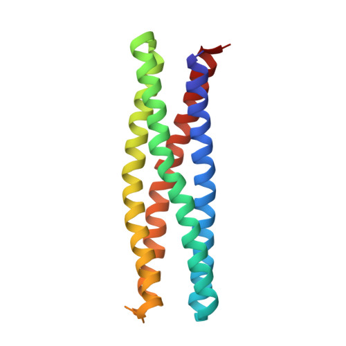
7JH6

De novo designed two-domain di-Zn(II) and porphyrin-binding protein


Experimental Data Snapshot

  • Method: X-RAY DIFFRACTION
  • Resolution: 3.50 Å
  • R-Value Free: 0.283 
  • R-Value Work: 0.261 
  • R-Value Observed: 0.262 

Starting Model: in silico
View more details

wwPDB Validation   3D Report Full Report


Ligand Structure Quality Assessment 


This is version 1.3 of the entry. See complete history


Literature

Allosteric cooperation in a de novo-designed two-domain protein.

Pirro, F.Schmidt, N.Lincoff, J.Widel, Z.X.Polizzi, N.F.Liu, L.Therien, M.J.Grabe, M.Chino, M.Lombardi, A.DeGrado, W.F.

(2020) Proc Natl Acad Sci U S A 117: 33246-33253

  • DOI: https://doi.org/10.1073/pnas.2017062117
  • Primary Citation of Related Structures:  
    7JH6

  • PubMed Abstract: 

    We describe the de novo design of an allosterically regulated protein, which comprises two tightly coupled domains. One domain is based on the DF (Due Ferri in Italian or two-iron in English) family of de novo proteins, which have a diiron cofactor that catalyzes a phenol oxidase reaction, while the second domain is based on PS1 (Porphyrin-binding Sequence), which binds a synthetic Zn-porphyrin (ZnP). The binding of ZnP to the original PS1 protein induces changes in structure and dynamics, which we expected to influence the catalytic rate of a fused DF domain when appropriately coupled. Both DF and PS1 are four-helix bundles, but they have distinct bundle architectures. To achieve tight coupling between the domains, they were connected by four helical linkers using a computational method to discover the most designable connections capable of spanning the two architectures. The resulting protein, DFP1 (Due Ferri Porphyrin), bound the two cofactors in the expected manner. The crystal structure of fully reconstituted DFP1 was also in excellent agreement with the design, and it showed the ZnP cofactor bound over 12 Å from the dimetal center. Next, a substrate-binding cleft leading to the diiron center was introduced into DFP1. The resulting protein acts as an allosterically modulated phenol oxidase. Its Michaelis-Menten parameters were strongly affected by the binding of ZnP, resulting in a fourfold tighter K m and a 7-fold decrease in k cat These studies establish the feasibility of designing allosterically regulated catalytic proteins, entirely from scratch.


  • Organizational Affiliation

    Department of Chemical Sciences, University of Napoli Federico II, 80126 Napoli, Italy.


Macromolecules
Find similar proteins by:  (by identity cutoff)  |  3D Structure
Entity ID: 1
MoleculeChains Sequence LengthOrganismDetailsImage
Two-domain di-Zn(II) and porphyrin-binding protein
A, B, C, D
174synthetic constructMutation(s): 0 
Entity Groups  
Sequence Clusters30% Identity50% Identity70% Identity90% Identity95% Identity100% Identity
Sequence Annotations
Expand
  • Reference Sequence
Small Molecules
Ligands 3 Unique
IDChains Name / Formula / InChI Key2D Diagram3D Interactions
7BU (Subject of Investigation/LOI)
Query on 7BU

Download Ideal Coordinates CCD File 
G [auth A],
K [auth B],
N [auth C],
Q [auth D]
[5,10,15,20-tetrakis(trifluoromethyl)porphyrinato(2-)-kappa~4~N~21~,N~22~,N~23~,N~24~]zinc
C24 H8 F12 N4 Zn
HVSFTYLFHSNRAZ-HQJDZOCDSA-N
2PE
Query on 2PE

Download Ideal Coordinates CCD File 
H [auth A]NONAETHYLENE GLYCOL
C18 H38 O10
YZUUTMGDONTGTN-UHFFFAOYSA-N
ZN (Subject of Investigation/LOI)
Query on ZN

Download Ideal Coordinates CCD File 
E [auth A]
F [auth A]
I [auth B]
J [auth B]
L [auth C]
E [auth A],
F [auth A],
I [auth B],
J [auth B],
L [auth C],
M [auth C],
O [auth D],
P [auth D]
ZINC ION
Zn
PTFCDOFLOPIGGS-UHFFFAOYSA-N
Experimental Data & Validation

Experimental Data

  • Method: X-RAY DIFFRACTION
  • Resolution: 3.50 Å
  • R-Value Free: 0.283 
  • R-Value Work: 0.261 
  • R-Value Observed: 0.262 
  • Space Group: C 1 2 1
Unit Cell:
Length ( Å )Angle ( ˚ )
a = 172.478α = 90
b = 27.825β = 117.16
c = 188.494γ = 90
Software Package:
Software NamePurpose
REFMACrefinement
XDSdata reduction
XSCALEdata scaling
PHASERphasing

Structure Validation

View Full Validation Report


Ligand Structure Quality Assessment 


Entry History & Funding Information

Deposition Data


Funding OrganizationLocationGrant Number
National Institutes of Health/National Institute of General Medical Sciences (NIH/NIGMS)United StatesR01GM117593

Revision History  (Full details and data files)

  • Version 1.0: 2020-12-09
    Type: Initial release
  • Version 1.1: 2021-06-23
    Changes: Database references
  • Version 1.2: 2024-03-06
    Changes: Data collection, Database references, Refinement description
  • Version 1.3: 2024-04-03
    Changes: Refinement description