


Crystal Structure of Leucine-, isoleucine-, valine-, threonine-, and alanine-binding protein from Brucella ovis, closed conformation
Abendroth, J., Dranow, D.M., Lorimer, D.D., Horanyi, P.S., Edwards, T.E.To be published.
Experimental Data Snapshot
Starting Model: experimental
View more details
wwPDB Validation 3D Report Full Report

Entity ID: 1 | |||||
|---|---|---|---|---|---|
| Molecule | Chains | Sequence Length | Organism | Details | Image |
| Putative branched chain amino acid ABC transporter, periplasmic amino acid-binding protein | 399 | Brucella ovis ATCC 25840 | Mutation(s): 0 Gene Names: BOV_A0021 | 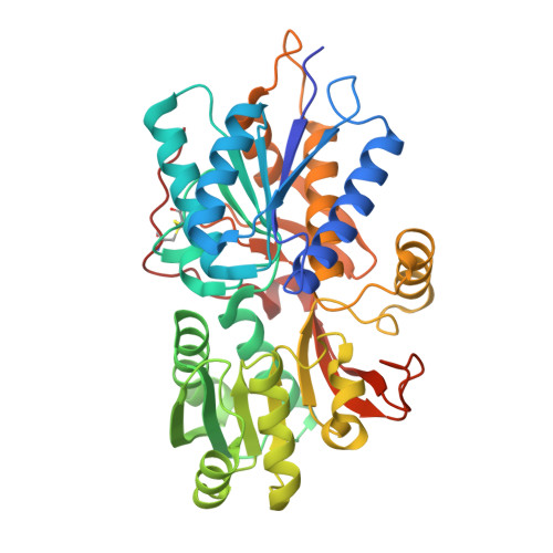 | |
UniProt | |||||
Find proteins for A0A0H3ATZ3 (Brucella ovis (strain ATCC 25840 / 63/290 / NCTC 10512)) Explore A0A0H3ATZ3 Go to UniProtKB: A0A0H3ATZ3 | |||||
Entity Groups | |||||
| Sequence Clusters | 30% Identity50% Identity70% Identity90% Identity95% Identity100% Identity | ||||
| UniProt Group | A0A0H3ATZ3 | ||||
Sequence AnnotationsExpand | |||||
Reference Sequence | |||||
| Ligands 3 Unique | |||||
|---|---|---|---|---|---|
| ID | Chains | Name / Formula / InChI Key | 2D Diagram | 3D Interactions | |
| EDO Download:Ideal Coordinates CCD File | F [auth A] | 1,2-ETHANEDIOL C2 H6 O2 LYCAIKOWRPUZTN-UHFFFAOYSA-N |  | ||
| NO3 Download:Ideal Coordinates CCD File | B [auth A], C [auth A], D [auth A] | NITRATE ION N O3 NHNBFGGVMKEFGY-UHFFFAOYSA-N |  | ||
| K Download:Ideal Coordinates CCD File | E [auth A] | POTASSIUM ION K NPYPAHLBTDXSSS-UHFFFAOYSA-N |  | ||
| Length ( Å ) | Angle ( ˚ ) |
|---|---|
| a = 46.77 | α = 90 |
| b = 69.39 | β = 90 |
| c = 120.75 | γ = 90 |
| Software Name | Purpose |
|---|---|
| XDS | data reduction |
| XSCALE | data scaling |
| PHENIX | refinement |
| PDB_EXTRACT | data extraction |
| PHASER | phasing |
| Coot | model building |


