A high-resolution data set of fatty acid-binding protein structures. III. Unexpectedly high occurrence of wrong ligands.
Ehler, A., Bartelmus, C., Benz, J., Plitzko, I., Rudolph, M.G.(2025) Acta Crystallogr D Struct Biol 81: 451-464
- PubMed: 40748031 Search on PubMedSearch on PubMed Central
- DOI: https://doi.org/10.1107/S2059798325006096
- Primary Citation Related Structures:
7FVU, 7FVV, 7FVW, 7FVX, 7FVY, 7FVZ, 7FW0, 7FW1, 7FW2, 7FW3, 7FW4, 7FW5, 7FW6, 7FW7, 7FW8, 7FW9, 7FWA, 7FWB, 7FWC, 7FWD, 7FWE, 7FWF, 7FWG, 7FWH, 7FWI, 7FWJ, 7FWK, 7FWL, 7FWM, 7FWN, 7FWO, 7FWP, 7FWQ, 7FWR, 7FWS, 7FWT, 7FWU, 7FWV, 7FWW, 7FWX, 7FWY, 7FWZ, 7FX0, 7FX1, 7FX2, 7FX3, 7FX4, 7FX5, 7FX6, 7FX7, ... Search all related entries - PubMed Abstract:
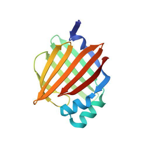
FABP4 has been implicated as a therapeutic target for treating diabetes and atherosclerosis. Structure-based drug design (SBDD) based on initial hits from high-throughput and fragment screens yielded 216 ligand-bound structures of human FABP3, FABP4 and FABP5 isoforms, many of which were at resolutions of better than 1.2 Å. An estimated 15% of the ligands had a different chemical composition to that expected from the starting materials or the final synthesis product, highlighting a potential problem inherent to all SBDD campaigns conducted at lower resolution. Apart from possible human error during compound registration, side reactions such as additions, eliminations, isomerizations, cyclizations and dimerizations were found that led to compounds capable of binding to FABP.
- Therapeutic Modalities, Innovation Center Basel, F. Hoffmann-La Roche, Grenzacherstrasse 124, 4070 Basel, Switzerland.
Organizational Affiliation: