7FIH

luteinizing hormone/choriogonadotropin receptor(S277I)-chorionic gonadotropin-Gs-Org43553 complex


Experimental Data Snapshot

  • Method: ELECTRON MICROSCOPY
  • Resolution: 3.20 Å
  • Aggregation State: PARTICLE 
  • Reconstruction Method: SINGLE PARTICLE 

wwPDB Validation   3D Report Full Report


This is version 1.1 of the entry. See complete history


Literature

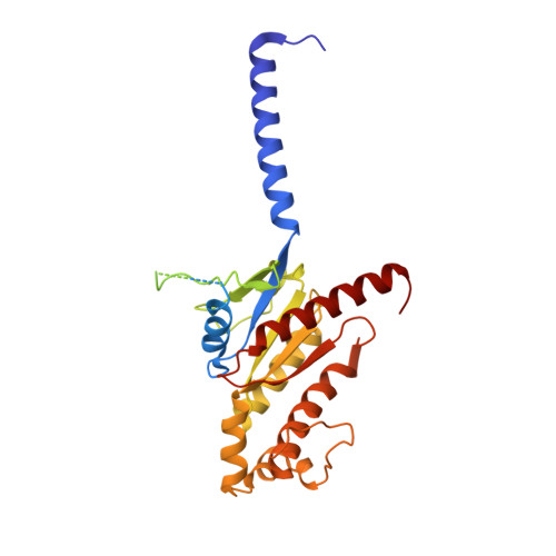
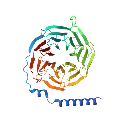
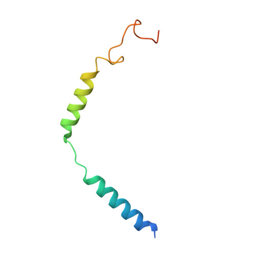
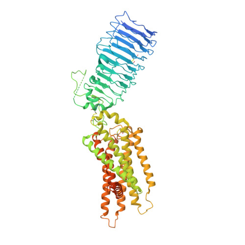
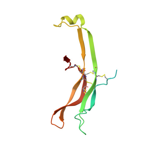
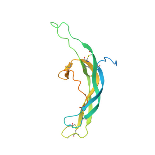
Structures of full-length glycoprotein hormone receptor signalling complexes.

Duan, J.Xu, P.Cheng, X.Mao, C.Croll, T.He, X.Shi, J.Luan, X.Yin, W.You, E.Liu, Q.Zhang, S.Jiang, H.Zhang, Y.Jiang, Y.Xu, H.E.

(2021) Nature 598: 688-692

  • DOI: https://doi.org/10.1038/s41586-021-03924-2
  • Primary Citation of Related Structures:  
    7FIG, 7FIH, 7FII, 7FIJ

  • PubMed Abstract: 

    Luteinizing hormone and chorionic gonadotropin are glycoprotein hormones that are related to follicle-stimulating hormone and thyroid-stimulating hormone 1,2 . Luteinizing hormone and chorionic gonadotropin are essential to human reproduction and are important therapeutic drugs 3-6 . They activate the same G-protein-coupled receptor, luteinizing hormone-choriogonadotropin receptor (LHCGR), by binding to the large extracellular domain 3 . Here we report four cryo-electron microscopy structures of LHCGR: two structures of the wild-type receptor in the inactive and active states; and two structures of the constitutively active mutated receptor. The active structures are bound to chorionic gonadotropin and the stimulatory G protein (G s ), and one of the structures also contains Org43553, an allosteric agonist 7 . The structures reveal a distinct 'push-and-pull' mechanism of receptor activation, in which the extracellular domain is pushed by the bound hormone and pulled by the extended hinge loop next to the transmembrane domain. A highly conserved 10-residue fragment (P10) from the hinge C-terminal loop at the interface between the extracellular domain and the transmembrane domain functions as a tethered agonist to induce conformational changes in the transmembrane domain and G-protein coupling. Org43553 binds to a pocket of the transmembrane domain and interacts directly with P10, which further stabilizes the active conformation. Together, these structures provide a common model for understanding the signalling of glycoprotein hormone receptors and a basis for drug discovery for endocrine diseases.


  • Organizational Affiliation

    The CAS Key Laboratory of Receptor Research and State Key Laboratory of Drug Research, Shanghai Institute of Materia Medica, Chinese Academy of Sciences, Shanghai, China.


Macromolecules
Find similar proteins by:  (by identity cutoff)  |  3D Structure
Entity ID: 1
MoleculeChains Sequence LengthOrganismDetailsImage
Engineered Guanine nucleotide-binding protein G(s) subunit alpha361Bos taurusMutation(s): 0 
Entity Groups  
Sequence Clusters30% Identity50% Identity70% Identity90% Identity95% Identity100% Identity
Sequence Annotations
Expand
  • Reference Sequence
Find similar proteins by:  (by identity cutoff)  |  3D Structure
Entity ID: 2
MoleculeChains Sequence LengthOrganismDetailsImage
Guanine nucleotide-binding protein G(I)/G(S)/G(T) subunit beta-1350Rattus norvegicusMutation(s): 0 
Gene Names: Gnb1
UniProt
Find proteins for P54311 (Rattus norvegicus)
Explore P54311 
Go to UniProtKB:  P54311
Entity Groups  
Sequence Clusters30% Identity50% Identity70% Identity90% Identity95% Identity100% Identity
UniProt GroupP54311
Sequence Annotations
Expand
  • Reference Sequence
Find similar proteins by:  (by identity cutoff)  |  3D Structure
Entity ID: 3
MoleculeChains Sequence LengthOrganismDetailsImage
Guanine nucleotide-binding protein G(I)/G(S)/G(O) subunit gamma-2C [auth G]71Bos taurusMutation(s): 0 
Gene Names: GNG2
Membrane Entity: Yes 
UniProt
Find proteins for P63212 (Bos taurus)
Explore P63212 
Go to UniProtKB:  P63212
Entity Groups  
Sequence Clusters30% Identity50% Identity70% Identity90% Identity95% Identity100% Identity
UniProt GroupP63212
Sequence Annotations
Expand
  • Reference Sequence
Find similar proteins by:  (by identity cutoff)  |  3D Structure
Entity ID: 4
MoleculeChains Sequence LengthOrganismDetailsImage
Lutropin-choriogonadotropic hormone receptorD [auth R]697Homo sapiensMutation(s): 1 
Gene Names: LHCGRLCGRLGR2LHRHR
Membrane Entity: Yes 
UniProt & NIH Common Fund Data Resources
Find proteins for P22888 (Homo sapiens)
Explore P22888 
Go to UniProtKB:  P22888
PHAROS:  P22888
GTEx:  ENSG00000138039 
Entity Groups  
Sequence Clusters30% Identity50% Identity70% Identity90% Identity95% Identity100% Identity
UniProt GroupP22888
Sequence Annotations
Expand
  • Reference Sequence
Find similar proteins by:  (by identity cutoff)  |  3D Structure
Entity ID: 5
MoleculeChains Sequence LengthOrganismDetailsImage
Glycoprotein hormones alpha chainE [auth X]115Homo sapiensMutation(s): 0 
Gene Names: CGA
Membrane Entity: Yes 
UniProt & NIH Common Fund Data Resources
Find proteins for P01215 (Homo sapiens)
Explore P01215 
Go to UniProtKB:  P01215
PHAROS:  P01215
GTEx:  ENSG00000135346 
Entity Groups  
Sequence Clusters30% Identity50% Identity70% Identity90% Identity95% Identity100% Identity
UniProt GroupP01215
Sequence Annotations
Expand
  • Reference Sequence
Find similar proteins by:  (by identity cutoff)  |  3D Structure
Entity ID: 6
MoleculeChains Sequence LengthOrganismDetailsImage
Choriogonadotropin subunit beta 3F [auth Y]165Homo sapiensMutation(s): 0 
Gene Names: CGB3CGBCGB5CGB8
Membrane Entity: Yes 
UniProt & NIH Common Fund Data Resources
Find proteins for P0DN86 (Homo sapiens)
Explore P0DN86 
Go to UniProtKB:  P0DN86
PHAROS:  P0DN86
Entity Groups  
Sequence Clusters30% Identity50% Identity70% Identity90% Identity95% Identity100% Identity
UniProt GroupP0DN86
Sequence Annotations
Expand
  • Reference Sequence
Small Molecules
Ligands 1 Unique
IDChains Name / Formula / InChI Key2D Diagram3D Interactions
55Z
Query on 55Z

Download Ideal Coordinates CCD File 
G [auth R]5-azanyl-N-tert-butyl-2-methylsulfanyl-4-[3-(2-morpholin-4-ylethanoylamino)phenyl]thieno[2,3-d]pyrimidine-6-carboxamide
C24 H30 N6 O3 S2
UWTXKCMCTINMHD-UHFFFAOYSA-N
Experimental Data & Validation

Experimental Data

  • Method: ELECTRON MICROSCOPY
  • Resolution: 3.20 Å
  • Aggregation State: PARTICLE 
  • Reconstruction Method: SINGLE PARTICLE 

Structure Validation

View Full Validation Report


Entry History & Funding Information

Deposition Data


Funding OrganizationLocationGrant Number
Ministry of Science and Technology (MoST, China)China2018YFA0507002

Revision History  (Full details and data files)

  • Version 1.0: 2021-09-29
    Type: Initial release
  • Version 1.1: 2022-02-16
    Changes: Database references