Surface Engineering of Top7 to Facilitate Structure Determination.
Ito, Y., Araki, T., Shiga, S., Konno, H., Makabe, K.(2022) Int J Mol Sci 23
- PubMed: 35054886 Search on PubMedSearch on PubMed Central
- DOI: https://doi.org/10.3390/ijms23020701
- Primary Citation Related Structures:
7FAO - PubMed Abstract:
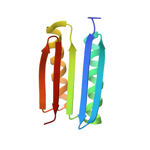
Top7 is a de novo designed protein whose amino acid sequence has no evolutional trace. Such a property makes Top7 a suitable scaffold for studying the pure nature of protein and protein engineering applications. To use Top7 as an engineering scaffold, we initially attempted structure determination and found that crystals of our construct, which lacked the terminal hexahistidine tag, showed weak diffraction in X-ray structure determination. Thus, we decided to introduce surface residue mutations to facilitate crystal structure determination. The resulting surface mutants, Top7sm1 and Top7sm2, crystallized easily and diffracted to the resolution around 1.7 Å. Despite the improved data, we could not finalize the structures due to high R values. Although we could not identify the origin of the high R values of the surface mutants, we found that all the structures shared common packing architecture with consecutive intermolecular β-sheet formation aligned in one direction. Thus, we mutated the intermolecular interface to disrupt the intermolecular β-sheet formation, expecting to form a new crystal packing. The resulting mutant, Top7sm2-I68R, formed new crystal packing interactions as intended and diffracted to the resolution of 1.4 Å. The surface mutations contributed to crystal packing and high resolution. We finalized the structure model with the R/R free values of 0.20/0.24. Top7sm2-I68R can be a useful model protein due to its convenient structure determination.
- Graduate School of Science and Engineering, Yamagata University, 4-3-16 Jyonan, Yonezawa 992-8510, Yamagata, Japan.
Organizational Affiliation: