7F81

Structure of the bacterial cellulose synthase subunit Z from Enterobacter sp. CJF-002


Experimental Data Snapshot

  • Method: X-RAY DIFFRACTION
  • Resolution: 1.93 Å
  • R-Value Free: 0.197 
  • R-Value Work: 0.162 
  • R-Value Observed: 0.164 

wwPDB Validation   3D Report Full Report


Ligand Structure Quality Assessment 


This is version 1.1 of the entry. See complete history


Literature

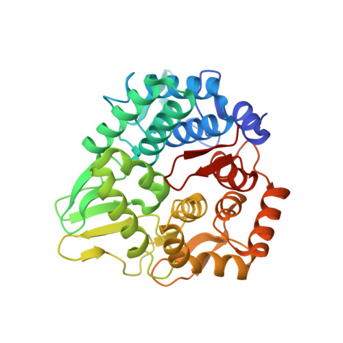
Structural snapshot of a glycoside hydrolase family 8 endo-beta-1,4-glucanase capturing the state after cleavage of the scissile bond.

Fujiwara, T.Fujishima, A.Nakamura, Y.Tajima, K.Yao, M.

(2022) Acta Crystallogr D Biol Crystallogr 78: 228-237


Macromolecules
Find similar proteins by:  (by identity cutoff)  |  3D Structure
Entity ID: 1
MoleculeChains Sequence LengthOrganismDetailsImage
Glucanase
A, B, C, D
351Enterobacter sp. CJF-002Mutation(s): 0 
Gene Names: bcsZ
EC: 3.2.1
UniProt
Find proteins for K0IUV6 (Enterobacter sp. CJF-002)
Explore K0IUV6 
Go to UniProtKB:  K0IUV6
Entity Groups  
Sequence Clusters30% Identity50% Identity70% Identity90% Identity95% Identity100% Identity
UniProt GroupK0IUV6
Sequence Annotations
Expand
  • Reference Sequence
Small Molecules
Ligands 2 Unique
IDChains Name / Formula / InChI Key2D Diagram3D Interactions
SRT
Query on SRT

Download Ideal Coordinates CCD File 
AA [auth D],
M [auth B]
S,R MESO-TARTARIC ACID
C4 H6 O6
FEWJPZIEWOKRBE-XIXRPRMCSA-N
GOL
Query on GOL

Download Ideal Coordinates CCD File 
E [auth A]
F [auth A]
G [auth B]
H [auth B]
I [auth B]
E [auth A],
F [auth A],
G [auth B],
H [auth B],
I [auth B],
J [auth B],
K [auth B],
L [auth B],
N [auth C],
O [auth C],
P [auth C],
Q [auth C],
R [auth C],
S [auth C],
T [auth D],
U [auth D],
V [auth D],
W [auth D],
X [auth D],
Y [auth D],
Z [auth D]
GLYCEROL
C3 H8 O3
PEDCQBHIVMGVHV-UHFFFAOYSA-N
Experimental Data & Validation

Experimental Data

  • Method: X-RAY DIFFRACTION
  • Resolution: 1.93 Å
  • R-Value Free: 0.197 
  • R-Value Work: 0.162 
  • R-Value Observed: 0.164 
  • Space Group: P 1 21 1
Unit Cell:
Length ( Å )Angle ( ˚ )
a = 89.267α = 90
b = 91.454β = 98.154
c = 89.931γ = 90
Software Package:
Software NamePurpose
PHENIXrefinement
PHENIXphasing
XDSdata scaling
XDSdata reduction

Structure Validation

View Full Validation Report


Ligand Structure Quality Assessment 


Entry History 

Deposition Data

Revision History  (Full details and data files)

  • Version 1.0: 2022-02-09
    Type: Initial release
  • Version 1.1: 2023-11-29
    Changes: Data collection, Refinement description