7F7F

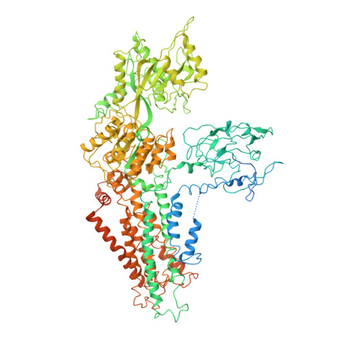
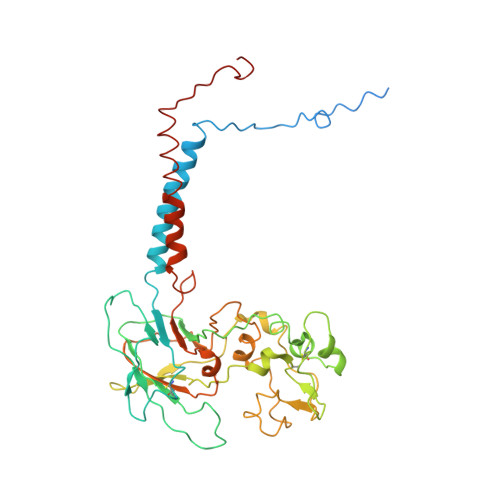
Cryo-EM structure of Dnf1 from Saccharomyces cerevisiae in yeast lipids with beryllium fluoride (resting state)


Experimental Data Snapshot

  • Method: ELECTRON MICROSCOPY
  • Resolution: 3.81 Å
  • Aggregation State: PARTICLE 
  • Reconstruction Method: SINGLE PARTICLE 

wwPDB Validation   3D Report Full Report


This is version 2.1 of the entry. See complete history


Literature

Conformational changes of a phosphatidylcholine flippase in lipid membranes.

Xu, J.He, Y.Wu, X.Li, L.

(2022) Cell Rep 38: 110518-110518

  • DOI: https://doi.org/10.1016/j.celrep.2022.110518
  • Primary Citation of Related Structures:  
    7DRX, 7DSH, 7DSI, 7F7F, 7WHV, 7WHW

  • PubMed Abstract: 

    Type 4 P-type ATPases (P4-ATPases) actively and selectively translocate phospholipids across membrane bilayers. Driven by ATP hydrolysis, P4-ATPases undergo conformational changes during lipid flipping. It is unclear how the active flipping states of P4-ATPases are regulated in the lipid membranes, especially for phosphatidylcholine (PC)-flipping P4-ATPases whose substrate, PC, is a substantial component of membranes. Here, we report the cryoelectron microscopy structures of a yeast PC-flipping P4-ATPase, Dnf1, in lipid environments. In native yeast lipids, Dnf1 adopts a conformation in which the lipid flipping pathway is disrupted. Only when the lipid composition is changed can Dnf1 be captured in the active conformations that enable lipid flipping. These results suggest that, in the native membrane, Dnf1 may stay in an idle conformation that is unable to support the trans-membrane movement of lipids. Dnf1 may have altered conformational preferences in membranes with different lipid compositions.


  • Organizational Affiliation

    State Key Laboratory of Membrane Biology, Peking-Tsinghua Center for Life Sciences, School of Life Sciences, Peking University, Beijing, China.


Macromolecules
Find similar proteins by:  (by identity cutoff)  |  3D Structure
Entity ID: 1
MoleculeChains Sequence LengthOrganismDetailsImage
Phospholipid-transporting ATPase DNF11,571Saccharomyces cerevisiae S288CMutation(s): 0 
Gene Names: DNF1YER166WSYGP-ORF7
EC: 7.6.2.1
Membrane Entity: Yes 
UniProt
Find proteins for P32660 (Saccharomyces cerevisiae (strain ATCC 204508 / S288c))
Explore P32660 
Go to UniProtKB:  P32660
Entity Groups  
Sequence Clusters30% Identity50% Identity70% Identity90% Identity95% Identity100% Identity
UniProt GroupP32660
Sequence Annotations
Expand
  • Reference Sequence
Find similar proteins by:  (by identity cutoff)  |  3D Structure
Entity ID: 2
MoleculeChains Sequence LengthOrganismDetailsImage
Alkylphosphocholine resistance protein LEM3414Saccharomyces cerevisiae S288CMutation(s): 0 
Gene Names: LEM3BRE3ROS3YNL323WN0333
Membrane Entity: Yes 
UniProt
Find proteins for P42838 (Saccharomyces cerevisiae (strain ATCC 204508 / S288c))
Explore P42838 
Go to UniProtKB:  P42838
Entity Groups  
Sequence Clusters30% Identity50% Identity70% Identity90% Identity95% Identity100% Identity
UniProt GroupP42838
Glycosylation
Glycosylation Sites: 3Go to GlyGen: P42838-1
Sequence Annotations
Expand
  • Reference Sequence
Oligosaccharides

Help

Entity ID: 3
MoleculeChains Length2D Diagram Glycosylation3D Interactions
2-acetamido-2-deoxy-beta-D-glucopyranose-(1-4)-2-acetamido-2-deoxy-beta-D-glucopyranose
C
2N-Glycosylation
Glycosylation Resources
GlyTouCan:  G42666HT
GlyCosmos:  G42666HT
GlyGen:  G42666HT
Entity ID: 4
MoleculeChains Length2D Diagram Glycosylation3D Interactions
beta-D-mannopyranose-(1-4)-2-acetamido-2-deoxy-beta-D-glucopyranose-(1-4)-2-acetamido-2-deoxy-beta-D-glucopyranose
D
3N-Glycosylation
Glycosylation Resources
GlyTouCan:  G15407YE
GlyCosmos:  G15407YE
GlyGen:  G15407YE
Entity ID: 5
MoleculeChains Length2D Diagram Glycosylation3D Interactions
2-acetamido-2-deoxy-beta-D-glucopyranose-(1-6)-2-acetamido-2-deoxy-beta-D-glucopyranose
E
2N-Glycosylation
Glycosylation Resources
GlyTouCan:  G35932AR
GlyCosmos:  G35932AR
GlyGen:  G35932AR
Small Molecules
Ligands 1 Unique
IDChains Name / Formula / InChI Key2D Diagram3D Interactions
MG
Query on MG

Download Ideal Coordinates CCD File 
F [auth A]MAGNESIUM ION
Mg
JLVVSXFLKOJNIY-UHFFFAOYSA-N
Experimental Data & Validation

Experimental Data

  • Method: ELECTRON MICROSCOPY
  • Resolution: 3.81 Å
  • Aggregation State: PARTICLE 
  • Reconstruction Method: SINGLE PARTICLE 

Structure Validation

View Full Validation Report


Entry History & Funding Information

Deposition Data


Funding OrganizationLocationGrant Number
National Natural Science Foundation of China (NSFC)China31870835

Revision History  (Full details and data files)

  • Version 1.0: 2022-03-23
    Type: Initial release
  • Version 1.1: 2022-03-30
    Changes: Database references
  • Version 2.0: 2022-04-20
    Type: Coordinate replacement
    Reason: Model orientation/position
    Changes: Advisory, Atomic model, Data collection, Derived calculations, Refinement description
  • Version 2.1: 2024-10-09
    Changes: Data collection, Structure summary