7F6J

Crystal structure of the PDZD8 coiled-coil domain - Rab7 complex


Experimental Data Snapshot

  • Method: X-RAY DIFFRACTION
  • Resolution: 2.10 Å
  • R-Value Free: 0.253 
  • R-Value Work: 0.207 
  • R-Value Observed: 0.209 

wwPDB Validation   3D Report Full Report


Ligand Structure Quality Assessment 


This is version 1.1 of the entry. See complete history


Literature

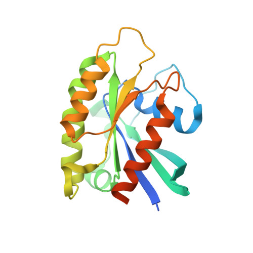
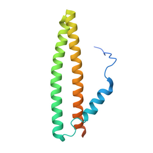
Structural basis of human PDZD8-Rab7 interaction for the ER-late endosome tethering.

Khan, H.Chen, L.Tan, L.Im, Y.J.

(2021) Sci Rep 11: 18859-18859

  • DOI: https://doi.org/10.1038/s41598-021-98419-5
  • Primary Citation of Related Structures:  
    7F6J

  • PubMed Abstract: 

    The membrane contact sites (MCSs) between the ER and late endosomes (LEs) are essential for the regulation of endosomal protein sorting, dynamics, and motility. PDZD8 is an ER transmembrane protein containing a Synaptotagmin-like Mitochondrial lipid-binding Proteins (SMP) domain. PDZD8 tethers the ER to late endosomes and lysosomes by associating its C-terminal coiled-coil (CC) with the LE Rab7. To identify the structural determinants for the PDZD8-Rab7 interaction, we determined the crystal structure of the human PDZD8 CC domain in complex with the GTP-bound form of Rab7. The PDZD8 CC contains one short helix and the two helices forming an antiparallel coiled-coil. Two Rab7 molecules bind to the opposite sides of the PDZD8 CC in a 2:1 ratio. The switch I/II and interswitch regions of the GTP-loaded Rab7 form the binding interfaces, which correlates with the GTP-dependent interaction of PDZD8 and Rab7. Analysis of the protein interaction by isothermal titration calorimetry confirms that two Rab7 molecules bind the PDZD8 CC in a GTP-dependent manner. The structural model of the PDZD8 CC-Rab7 complex correlates with the recruitment of PDZD8 at the LE-ER interface and its role in lipid transport and regulation.


  • Organizational Affiliation

    College of Pharmacy, Chonnam National University, Gwangju, 61186, Republic of Korea.


Macromolecules
Find similar proteins by:  (by identity cutoff)  |  3D Structure
Entity ID: 1
MoleculeChains Sequence LengthOrganismDetailsImage
Ras-related protein Rab-7a
A, B
200Homo sapiensMutation(s): 1 
Gene Names: RAB7ARAB7
EC: 3.6.5.2
UniProt & NIH Common Fund Data Resources
Find proteins for P51149 (Homo sapiens)
Explore P51149 
Go to UniProtKB:  P51149
PHAROS:  P51149
GTEx:  ENSG00000075785 
Entity Groups  
Sequence Clusters30% Identity50% Identity70% Identity90% Identity95% Identity100% Identity
UniProt GroupP51149
Sequence Annotations
Expand
  • Reference Sequence
Find similar proteins by:  (by identity cutoff)  |  3D Structure
Entity ID: 2
MoleculeChains Sequence LengthOrganismDetailsImage
PDZ domain-containing protein 8133Homo sapiensMutation(s): 0 
Gene Names: PDZD8PDZK8
UniProt & NIH Common Fund Data Resources
Find proteins for Q8NEN9 (Homo sapiens)
Explore Q8NEN9 
Go to UniProtKB:  Q8NEN9
PHAROS:  Q8NEN9
GTEx:  ENSG00000165650 
Entity Groups  
Sequence Clusters30% Identity50% Identity70% Identity90% Identity95% Identity100% Identity
UniProt GroupQ8NEN9
Sequence Annotations
Expand
  • Reference Sequence
Experimental Data & Validation

Experimental Data

  • Method: X-RAY DIFFRACTION
  • Resolution: 2.10 Å
  • R-Value Free: 0.253 
  • R-Value Work: 0.207 
  • R-Value Observed: 0.209 
  • Space Group: P 65
Unit Cell:
Length ( Å )Angle ( ˚ )
a = 84.226α = 90
b = 84.226β = 90
c = 154.622γ = 120
Software Package:
Software NamePurpose
ADSCdata collection
HKL-2000data reduction
HKL-2000data scaling
PHASERphasing
PHENIXrefinement

Structure Validation

View Full Validation Report


Ligand Structure Quality Assessment 


Entry History & Funding Information

Deposition Data


Funding OrganizationLocationGrant Number
National Research Foundation (NRF, Korea)Korea, Republic Of2019R1A2C1085530

Revision History  (Full details and data files)

  • Version 1.0: 2022-02-23
    Type: Initial release
  • Version 1.1: 2023-11-29
    Changes: Data collection, Refinement description